Anti-CYP1A2 Antibody (clone 2E9)
| Name | Anti-CYP1A2 Antibody (clone 2E9) |
|---|---|
| Supplier | LifeSpan Bioscience |
| Catalog | LS-C115226 |
| Host | Mouse |
| Clonality | Monoclonal |
| Isotype | IgG1 |
| Clone | 2E9 |
| Applications | WB FC |
| Species Reactivities | Human |
| Antigen | CYP1A2 antibody was raised against full length human recombinant protein of human CYP1A2 (NP_000752) produced in HEK293T cell |
| Description | Mouse Monoclonal |
| Gene | CYP1A2 |
| Conjugate | Unconjugated |
| Supplier Page | Shop |
Product images
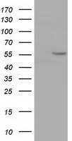 HEK293T cells were transfected with the pCMV6-ENTRY control (Left lane) or pCMV6-ENTRY CYP1A2 (Right lane) cDNA for 48 hrs and lysed. Equivalent amounts of cell lysates (5 ug per lane) were separated by SDS-PAGE and immunoblotted with anti-CYP1A2.
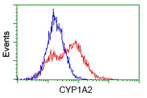
 Flow cytometry of Jurkat cells, using anti-CYP1A2 antibody, (Red), compared to a nonspecific negative control antibody, (Blue).
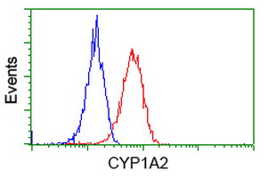 Flow cytometry of HeLa cells, using anti-CYP1A2 antibody, (Red), compared to a nonspecific negative control antibody, (Blue).
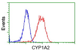
 HEK293T cells transfected with either overexpress plasmid (Red) or empty vector control plasmid (Blue) were immunostained by anti-CYP1A2 antibody, and then analyzed by flow cytometry.