Anti-REM 2 (GTP-binding protein) Antibody
| Name | Anti-REM 2 (GTP-binding protein) Antibody |
|---|---|
| Supplier | Sigma-Aldrich |
| Catalog | ABD37 |
| Prices | $309.00 |
| Sizes | abd37 |
| Clonality | Polyclonal |
| Applications | WB |
| Species Reactivities | Human, Mouse |
| Purity/Format | Affinity Purfied |
| Description | Polyclonal |
| Gene | REM2 |
| Supplier Page | Shop |
Product images
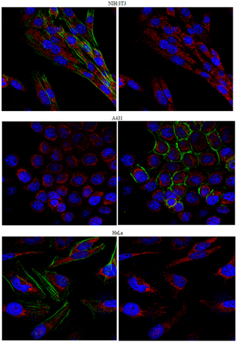 Immunocytochemistry Analysis:
 Representative lot data.
Confocal fluorescent analysis of HeLa, A431, and NIH/3T3 cells using Cat. No. ABD37, Anti-REM 2 (GTP-binding protein) (1:500 dilution) and a Donkey Anti-Rabbit IgG secondary antibody conjugated to Cy3 (Red). Actin filaments have been labeled with Alexa Fluor® 488 dye - Phalloidin (Green). Nucleus is stained with DAPI (Blue). This antibody positively stains the cytoplasm.
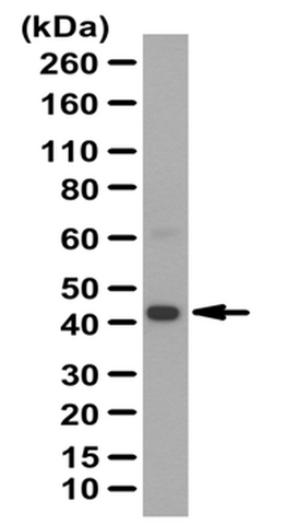 Western Blotting Analysis:
 Representative lot data.
Human kidney tissue lysate was probed with Cat. No. ABD37, Anti-REM 2 (GTP-binding protein) (0.5 µg/mL).
Proteins were visualized using a Donkey Anti-Rabbit IgG secondary antibody conjugated to HRP and a chemiluminescence detection system.
Arrow indicates REM2 (~42 kDa). Note: An uncharacterized band appears at ~62 kDa in some lysates.
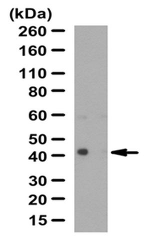 Western Blotting Analysis:
 Representative lot data.
Human heart tissue lysate was probed with Cat. No. ABD37, Anti-REM 2 (GTP-binding protein) (0.5 µg/mL).
Proteins were visualized using a Donkey Anti-Rabbit IgG secondary antibody conjugated to HRP and a chemiluminescence detection system.
Arrow indicates REM2 (~42 kDa). Note: An uncharacterized band appears at ~62 kDa in some lysates.