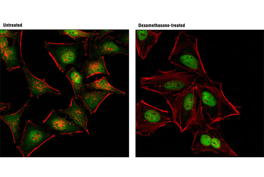
Confocal immunofluorescent analysis of HeLa cells, grown in phenol red-free media containing 5% charcoal-stripped FBS for 2 d, and either untreated (left) or dexamethasone-treated (100 nM, 2 hr; right), using Glucocorticoid Receptor (D8H2) XP ® Rabbit mAb (Alexa Fluor ® 488 Conjugate) (green). Actin filaments were labeled with DY-554 phalloidin (red).

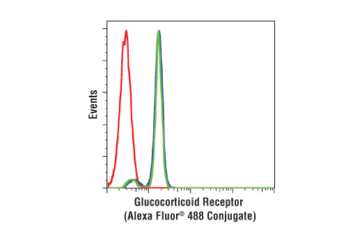
Human whole blood was fixed, lysed, and permeabilized as per the Cell Signaling Technology Flow Cytometry (Alternate) Protocol and stained with CD3-PE, CD19-APC and Glucocorticoid Receptor (D8H2) XP® Rabbit mAb (Alexa Fluor® 488 Conjugate). CD3 (blue) and CD19 (green) population gates were applied to a histogram depicting the mean fluorescence intensity of glucocorticoid and compared to Rabbit (DA1E) mAb IgG XP® Isotype Control (Alexa Fluor ® 488 Conjugate) #2975 (red).

