Anti-DLD / Diaphorase / E3 Antibody (clone 8A10)
| Name | Anti-DLD / Diaphorase / E3 Antibody (clone 8A10) |
|---|---|
| Supplier | LifeSpan Bioscience |
| Catalog | LS-C173227 |
| Prices | $325.00 |
| Sizes | 100 µl |
| Host | Mouse |
| Clonality | Monoclonal |
| Isotype | IgG1 |
| Clone | 8A10 |
| Applications | IHC-P ICC/IF WB IP ELISA |
| Species Reactivities | Pig, Human |
| Antigen | Full length human recombinant protein of human DLD(NP_000099) produced in HEK293T cell. |
| Purity/Format | Protein A/G purified |
| Description | Mouse Monoclonal |
| Gene | DLD |
| Conjugate | Unconjugated |
| Supplier Page | Shop |
Product images
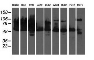 Western blot of extracts (35 ug) from 9 different cell lines by using anti-DLD monoclonal antibody (HepG2: human; HeLa: human; SVT2: mouse; A549: human; COS7: monkey; Jurkat: human; MDCK: canine; PC12: rat; MCF7: human).
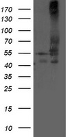 HEK293T cells were transfected with the pCMV6-ENTRY control (Left lane) or pCMV6-ENTRY DLD (Right lane) cDNA for 48 hrs and lysed. Equivalent amounts of cell lysates (5 ug per lane) were separated by SDS-PAGE and immunoblotted with anti-DLD.
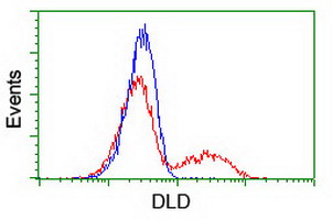 HEK293T cells transfected with either overexpress plasmid (Red) or empty vector control plasmid (Blue) were immunostained by anti-DLD antibody, and then analyzed by flow cytometry.