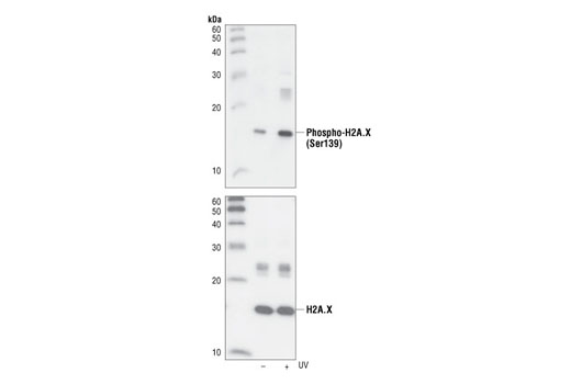
Western blot analysis of extracts from untreated or UV-treated 293 cells, using Phospho-Histone H2A.X (Ser139) (20E3) Rabbit mAb (upper) or Histone H2A.X Antibody #2595 (lower).

Immunohistochemical analysis of paraffin-embedded HT-29 cells untreated (left) or UV-treated (right), using Phospho-Histone H2A.X (Ser139) (20E3) Rabbit mAb.

Immunohistochemical analysis of paraffin-embedded human breast carcinoma, using Phospho-Histone H2A.X (Ser139) (20E3) Rabbit mAb in the presence of control peptide (left) or Phospho-Histone H2A.X (Ser139) Blocking Peptide #1260 (right).

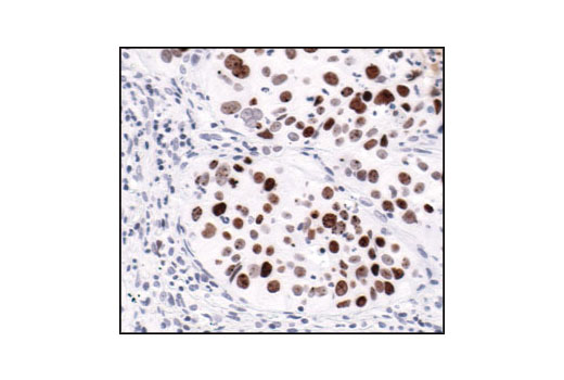
Immunohistochemical analysis of paraffin-embedded human colon carcinoma, using Phospho-Histone H2A.X (Ser139) (20E3) Rabbit mAb.

Immunohistochemical analysis of paraffin-embedded human lung carcinoma, using Phospho-Histone H2A.X (Ser139) (20E3) Rabbit mAb, showing nuclear localization.

Immunohistochemical analysis of paraffin-embedded human lung carcinoma untreated (left) or lambda-phosphatase-treated (right), using Phospho-Histone H2A.X (Ser139) (20E3) Rabbit mAb.

Confocal immunofluorescent analysis of HeLa cells, untreated (left) or UV-treated (right), using Phospho-Histone H2A.X (Ser139) (20E3) Rabbit mAb (green). Actin filaments have been labeled with DY-554 phalloidin (red).

Flow cytometric analysis of HeLa cells, untreated (blue) or UV-treated (green), using Phospho-Histone H2A.X (Ser139) (20E3) Rabbit mAb compared to a nonspecific negative control antibody (red).


