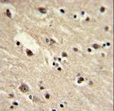
Formalin-fixed and paraffin-embedded human brain with EEF1A1 Antibody , which was peroxidase-conjugated to the secondary antibody, followed by DAB staining. This data demonstrates the use of this antibody for immunohistochemistry; clinical relevance has not been evaluated.

With 293T cell line lysate,resolved proteins were electrophoretically transferred to PVDF membrane and incubated sequentially with primary antibody EF1A1(1:500, 4 degrees C overnight ) and horseradish peroxidase-conjugated second antibody (rabbit). After washing, the bound antibody complex was detected using an ECL chemiluminescence reagent and XAR film (Kodak).

Western blot of EEF1A1 Antibody in Y79, T47D cell line lysates (35 ug/lane). EEF1A1 (arrow) was detected using the purified antibody.

Flow cytometric of NCI-H292 cells using EEF1A1 Antibody (bottom histogram) compared to a negative control cell (top histogram). FITC-conjugated goat-anti-rabbit secondary antibodies were used for the analysis.


