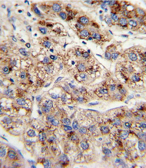
Formalin-fixed and paraffin-embedded human hepatocarcinoma with EHHADH Antibody , which was peroxidase-conjugated to the secondary antibody, followed by DAB staining. This data demonstrates the use of this antibody for immunohistochemistry; clinical relevance has not been evaluated.

Western blot of EHHADH Antibody in mouse liver (lane 1), kidney(lane 2) tissue lysates (35 ug/lane). EHHADH (arrow) was detected using the purified antibody.

Western blot of lysates from mouse kidney, rat kidney and liver tissue (from left to right), using EHHADH Antibody. Antibody was diluted at 1:1000 at each lane. A goat anti-rabbit IgG H&L (HRP) at 1:5000 dilution was used as the secondary antibody. Lysates at 35ug per lane.

Flow cytometric of HepG2 cells using EHHADH Antibody (bottom histogram) compared to a negative control cell (top histogram). FITC-conjugated goat-anti-rabbit secondary antibodies were used for the analysis.