
ARG55696 anti-Glutaminase antibody WB image Western blot: 35 µg of Mouse liver tissue lysates stained with ARG55696 anti-Glutaminase antibody.
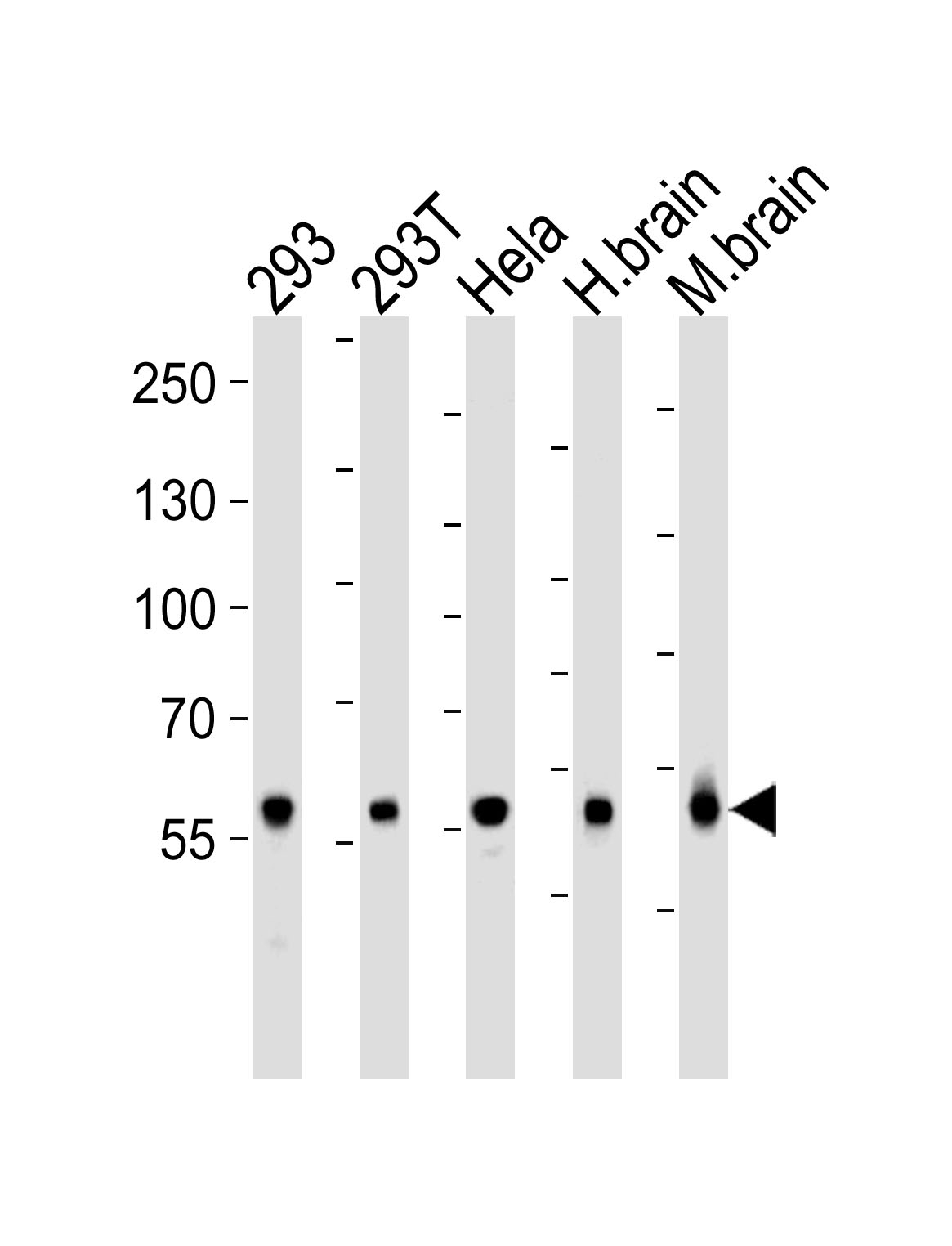
ARG55696 anti-Glutaminase antibody WB image Western blot: 35 µg of 293, 293T, HeLa cell line , huamn brain and Mouse brain tissue lysate (from left to right) stained with ARG55696 anti-Glutaminase antibody at 1:1000 dilution.
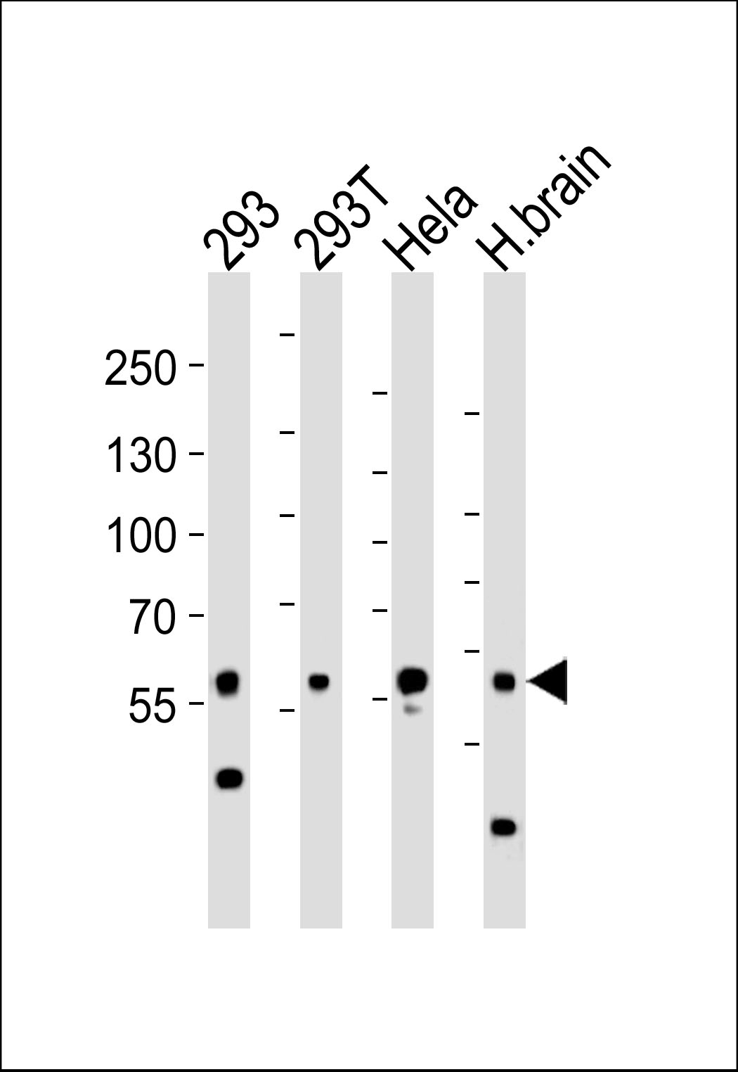
ARG55696 anti-Glutaminase antibody WB image Western blot: 35 µg of 293, 293T, HeLa cell line and Human brain tissue lysate (from left to right) stained with ARG55696 anti-Glutaminase antibody at 1:1000 dilution.
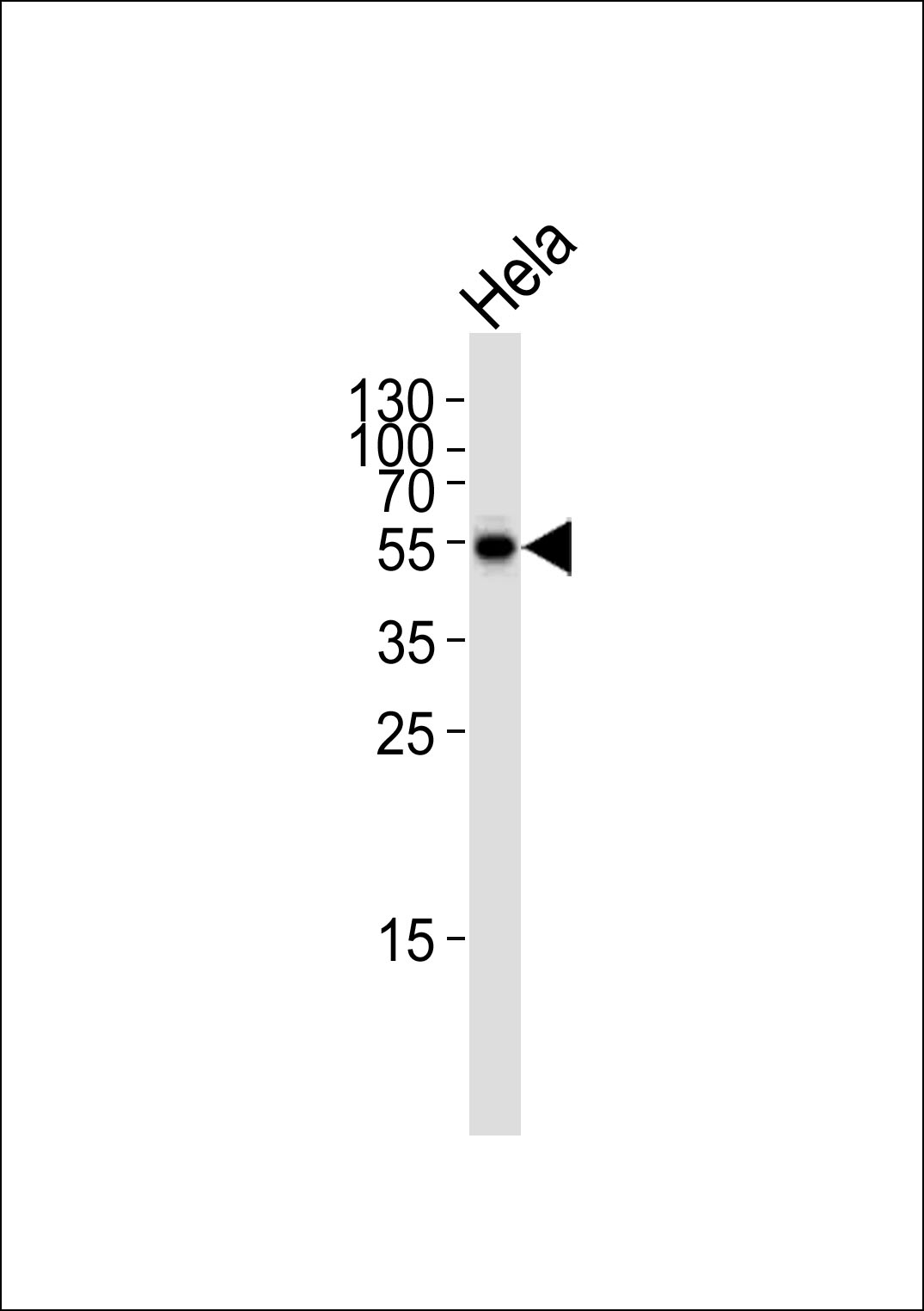
ARG55696 anti-Glutaminase antibody WB image Western blot: 35 µg of HeLa cell line stained with ARG55696 anti-Glutaminase antibody at 1:1000 dilution.
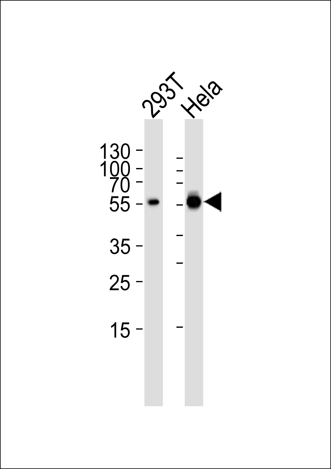
ARG55696 anti-Glutaminase antibody WB image Western blot: 35 µg of 293T, HeLa cell line (from left to right) stained with ARG55696 anti-Glutaminase antibody at 1:1000 dilution.
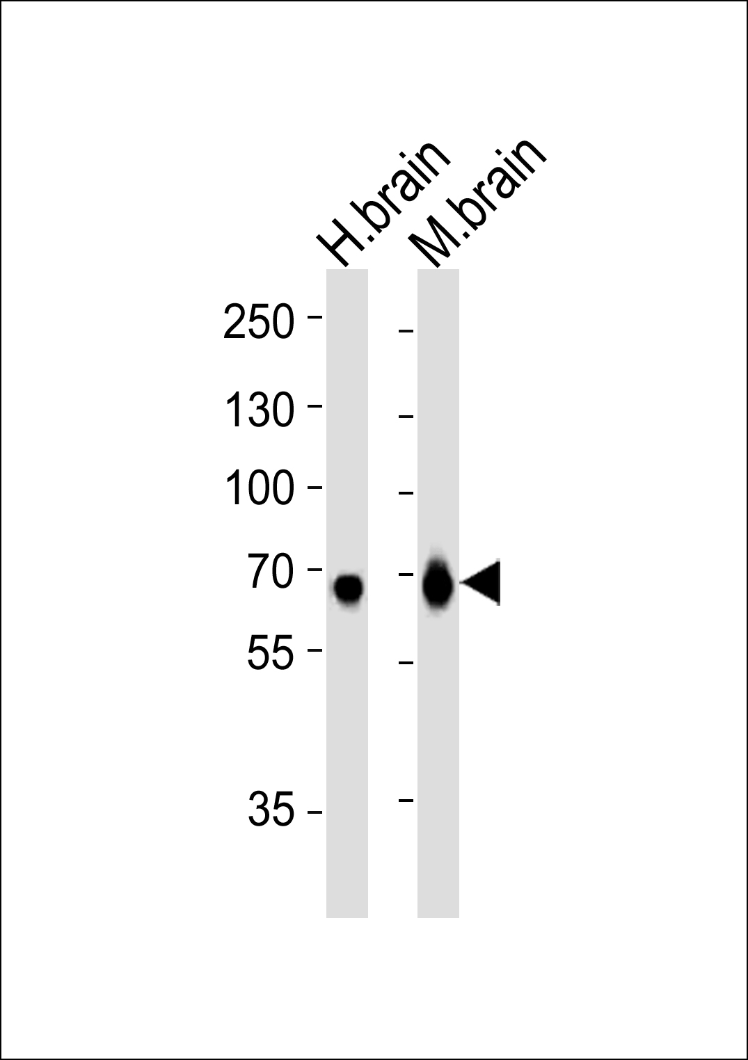
ARG55696 anti-Glutaminase antibody WB image Western blot: 35 µg of Human brain and Mouse brain tissue lysate (from left to right) stained with ARG55696 anti-Glutaminase antibody at 1:1000 dilution.
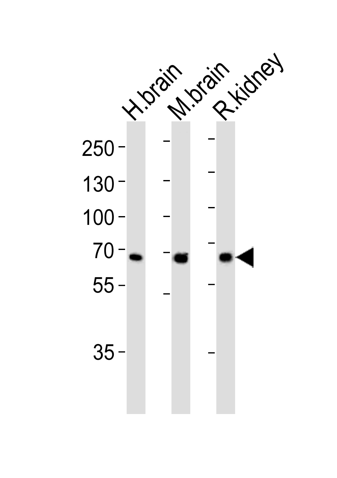
ARG55696 anti-Glutaminase antibody WB image Western blot: 35 µg of Human brain, Mouse brain ad Rat kidney tissue lysate (from left to right) stained with ARG55696 anti-Glutaminase antibody at 1:1000 dilution.
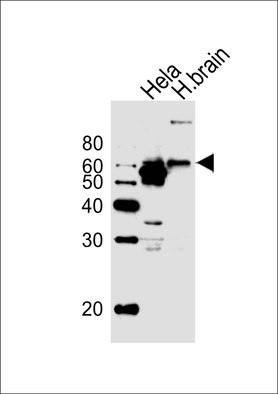
ARG55696 anti-Glutaminase antibody WB image Western blot: 20 µg of Human brain tissue and HeLa cell line (from left to right) stained with ARG55696 anti-Glutaminase antibody at 1:1000 dilution.
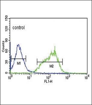
ARG55696 anti-Glutaminase antibody FACS image Flow Cytometry: HepG2 cells stained with ARG55696 anti-Glutaminase antibody (right histogram) or without primary antibody control (left histogram), followed by incubation with FITC labelled secondary antibody.
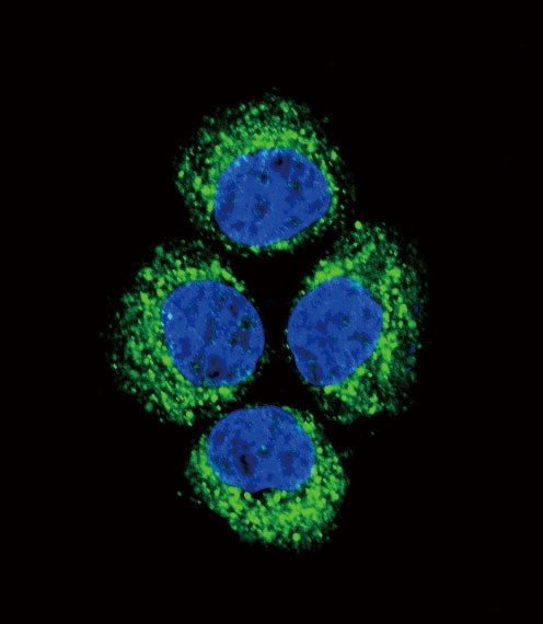
ARG55696 anti-Glutaminase antibody ICC/IF image Immunofluorescence: HeLa cell stained with ARG55696 anti-Glutaminase antibody (green). DAPI was used to stain the cell nuclear (blue).
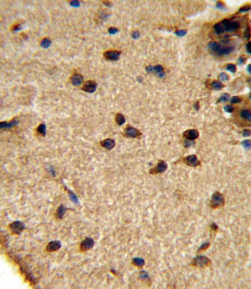
ARG55696 anti-Glutaminase antibody IHC-P image Immunohistochemistry: formalin fixed and paraffin embedded Mouse brain tissue stained with ARG55696 anti-Glutaminase antibody.
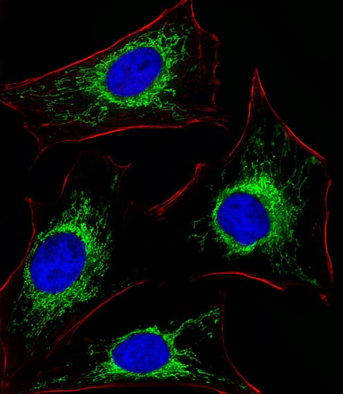
ARG55696 anti-Glutaminase antibody ICC/IF image Immunofluorescence: HeLa cells stained with ARG55696 anti-Glutaminase antibody (green) at 1:25 dilution. DAPI was used to stain the cell nuclear (blue). Cytoplasmic actin was counterstained with Alexa Fluor® 555 conjugated with Phalloidin (red).
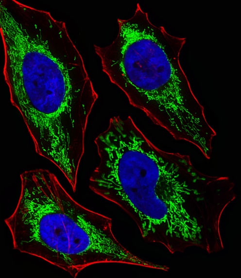
ARG55696 anti-Glutaminase antibody ICC/IF image Immunofluorescence: HeLa cells stained with ARG55696 anti-Glutaminase antibody (green) at 1:25 dilution. DAPI was used to stain the cell nuclear (blue). Cytoplasmic actin was counterstained with Alexa Fluor® 555 conjugated with Phalloidin (red).
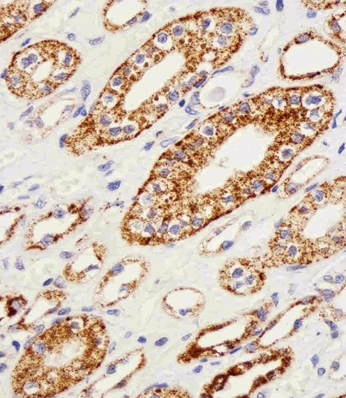
ARG55696 anti-Glutaminase antibody IHC-P image Immunohistochemistry: paraffin-embedded H.kidney section stained with ARG55696 anti-Glutaminase antibody at 1:25 dilution.