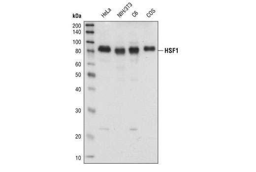
Western blot analysis of extracts from HeLa, NIH/3T3, C6 and COS cells, using HSF1 antibody.

Immunohistochemical analysis of paraffin-embedded human breast carcinoma, using HSF1 Antibody in the presence of control peptide (left) or antigen-specific peptide (right).
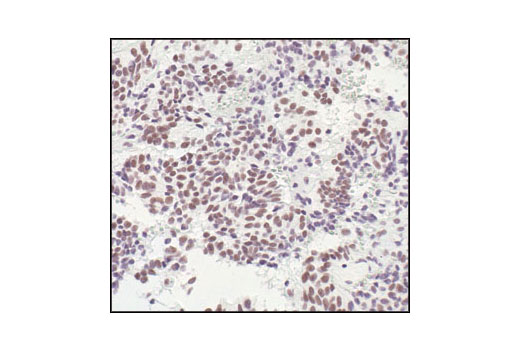
Immunohistochemical analysis of paraffin-embedded human pituitary adenoma, using HSF1 Antibody.

Immunohistochemical analysis of paraffin-embedded human breast carcinoma showing nuclear localization, using HSF1 Antibody.

Immunohistochemical analysis of paraffin-embedded human colon carcinoma, using HSF1 Antibody.

Immunohistochemical analysis of paraffin-embedded human lung carcinoma, using HSF1 Antibody.
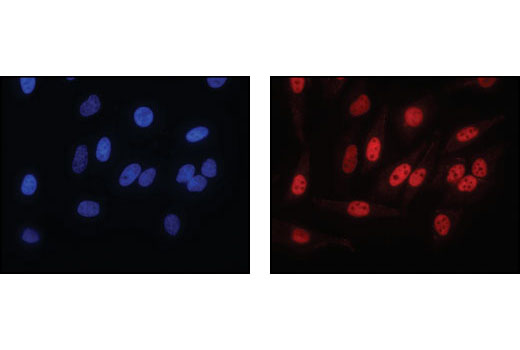
DAPI staining (left) and immunofluorescent staining (right) of paraformaldehyde-fixed HeLa cells, using HSF1 antibody.

Flow cytometric analysis of K562 cells, using HSF1 Antibody (blue) compared to a nonspecific negative control antibody (red).

HeLa cells were either untreated (left panel) or heat shocked (right panel) for 1h. Chromatin immunoprecipitations were performed with cross-linked chromatin from 4 x 10 6 cells and either 10 µl of HSF1 Antibody or 2 µl of Normal Rabbit IgG #2729 using SimpleChIP ® Enzymatic Chromatin IP Kit (Magnetic Beads) #9003. The enriched DNA was quantified by real-time PCR using SimpleChIP ® Human HSPA6 Promoter Primers #5551, human HSP70 intron 1 primers, and SimpleChIP ® Human α Satellite Repeat Primers #4486. The amount of immunoprecipitated DNA in each sample is represented as signal relative to the total amount of input chromatin, which is equivalent to one.