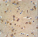
GABRG3 Antibody IHC of formalin-fixed and paraffin-embedded brain tissue followed by peroxidase-conjugated secondary antibody and DAB staining.

Western blot of GABRG3 Antibody in mouse brain tissue lysates (35 ug/lane). GABRG3 (arrow) was detected using the purified antibody.

Western blot of lysates from K562 cell line and human brain, testis tissue lysate(from left to right), using GABRG3 Antibody. Antibody was diluted at 1:1000 at each lane. A goat anti-rabbit IgG H&L (HRP) at 1:5000 dilution was used as the secondary antibody. Lysates at 35ug per lane.

GABRG3 Antibody flow cytometry of K562 cells (bottom histogram) compared to a negative control cell (top histogram). FITC-conjugated goat-anti-rabbit secondary antibodies were used for the analysis.