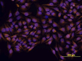
GLI-3 in HeLa Human Cell Line. GLI-3 was detected in immersion fixed HeLa human cervical epithelial carcinoma cell line using Human/Mouse GLI-3 Antigen Affinity-purified Polyclonal Antibody at 10 ug/ml for 3 hours at room temperature. Cells were stained using the NorthernLights 557-conjugated Anti-Goat IgG Secondary Antibody (yellow) and counterstained with DAPI (blue).
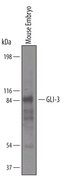
Detection of Mouse GLI3 by Western Blot. Western blot shows lysates of mouse embryo tissue. PVDF membrane was probed with 1 ug/ml of Goat Anti-Human/ Mouse GLI3 Antigen Affinity-purified Polyclonal Antibody followed by HRP-conjugated Anti-Goat IgG Secondary Antibody. A specific band was detected for GLI 3 at approximately 85 kD (as indicated). This experiment was conducted under reducing conditions.
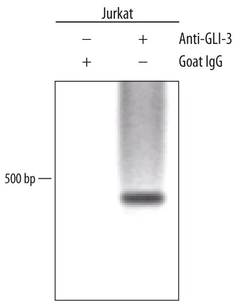
Detection of GLI-3-regulated Genes by Chromatin Immunoprecipitation. Jurkat human acute T cell leukemia cell line treated with 50 ng/ml PMA and 200 ng/ml calcium ionomycin for 30 minutes was fixed using formaldehyde, resuspended in lysis buffer, and sonicated to shear chromatin. GLI-3/DNA complexes were immunoprecipitated using 5 ug Goat Anti-Human/Mouse GLI-3 Antigen Affinity-purified Polyclonal Antibody or control antibody for 15 minutes in an ultrasonic bath, followed by Biotinylated Anti-Goat IgG Secondary Antibody. immune complexes were captured using 50 ul of MagCellect Streptavidin Ferrofluid and DNA was purified using chelating resin solution. The GLI-1 promoter was detected by standard PCR.