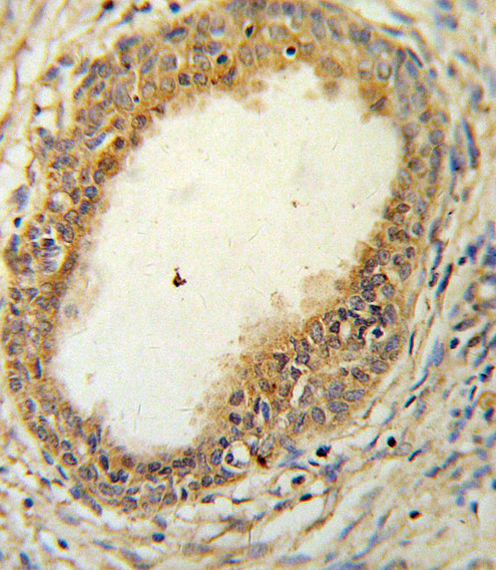
HAPLN1 Antibody IHC of formalin-fixed and paraffin-embedded prostate carcinoma followed by peroxidase-conjugated secondary antibody and DAB staining. This data demonstrates the use of the HAPLN1 Antibody for immunohistochemistry.
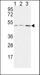
HAPLN1 Antibody western blot of MCF-7(lane 1),Jurkat(lane 2),HepG2(lane 3) cell line lysates (35 ug/lane). The HAPLN1 antibody detected the HAPLN1 protein (arrow).
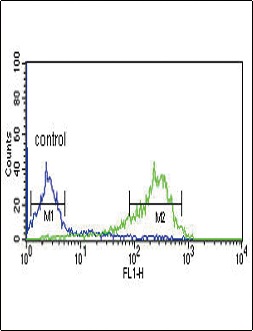
HAPLN1 Antibody flow cytometry of MCF-7 cells (right histogram) compared to a negative control cell (left histogram). FITC-conjugated goat-anti-rabbit secondary antibodies were used for the analysis.