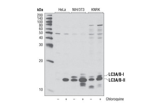
Western blot analysis of extracts from various cell lines, untreated or treated with chloroquine (50 μM, overnight) using LC3A/B Antibody.

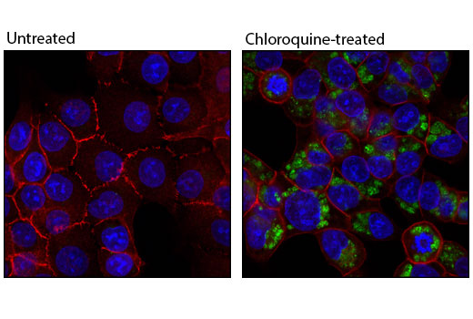
Confocal immunofluorescent analysis of HCT-116 cells, untreated (left) or choroquine-treated (50 uM, overnight; right) using LC3A/B Antibody (green) and β-Catenin (L54E2) Mouse mAb (Alexa Fluor® 555 Conjugate) #5612 (red). Blue pseudocolor = DRAQ5® #4084 (fluorescent DNA dye).

Flow cytometric analysis of Hela cells using LC3A/B Antibody (blue) compared to a nonspecific negative control antibody (red).


