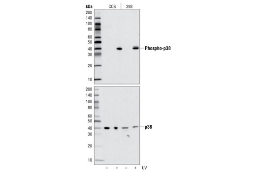
Western blot analysis of extracts from COS and 293 cells, untreated or UV-treated, using Phospho-p38 MAPK (Thr180/Tyr182) (D3F9) XP ® Rabbit mAb (upper) or p38 MAPK Antibody #9212 (lower).
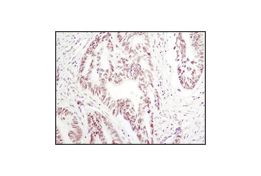
Immunohistochemical analysis of paraffin-embedded human colon carcinoma using Phospho-p38 MAPK (Thr180/Tyr182) (D3F9) XP ® Rabbit mAb.
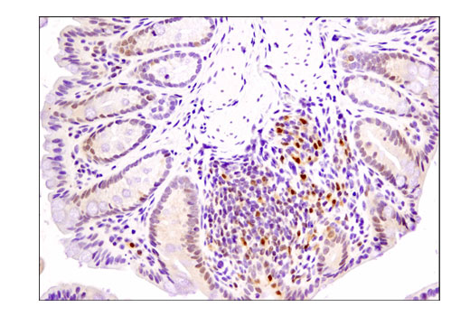
Immunohistochemical analysis of paraffin-embedded mouse colon using Phospho-p38 MAPK (Thr180/Tyr182) (D3F9) XP® Rabbit mAb.
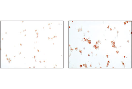
Immunohistochemical analysis of paraffin-embedded 293T cell pellets, untreated (left) or UV-treated (right), using Phospho-p38 MAPK (Thr180/Tyr182) (D3F9) XP ® Rabbit mAb.
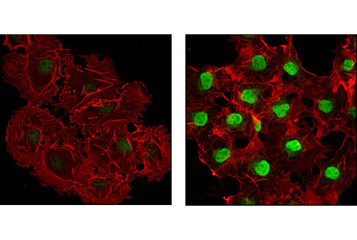
Confocal immunofluorescent analysis of COS cells, untreated (left) or anisomycin-treated (right) using Phospho-p38 MAPK (Thr180/Tyr182) (D3F9) XP ® Rabbit mAb (green). Actin filaments have been labeled with DY-554 phalloidin (red).
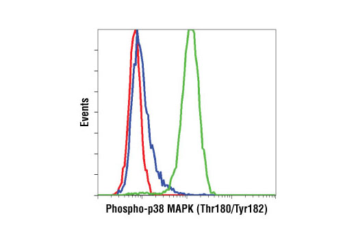
Flow cytometric analysis of Jurkat cells, untreated (blue) or anisomycin-treated (green), using Phospho-p38 MAPK (Thr180/Tyr182) (D3F9) XP ® Rabbit mAb compared to a nonspecific negative control antibody (red).


