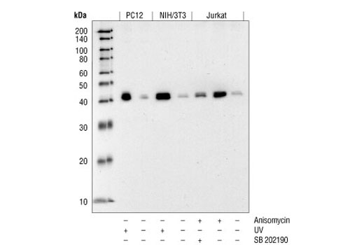
Western blot analysis of extracts from Jurkat, NIH/3T3 and PC12 cells, untreated or treated as indicated, using Phospho-p38 MAPK (Thr180/Tyr182) (12F8) Rabbit mAb.

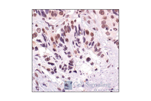
Immunohistochemistry of paraffin-embedded lung carcinoma, showing nuclear localization, using Phospho-p38 MAPK (Thr180/Tyr182) (12F8) Rabbit mAb.

Immunohistochemistry of paraffin-embedded colon carcinoma, showing nuclear localization, using Phospho-p38 MAPK (Thr180/Tyr182) (12F8) Rabbit mAb.

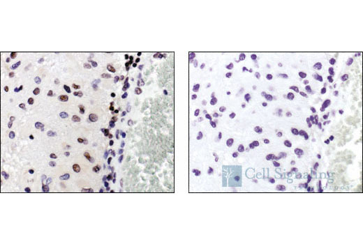
Immunohistochemistry of paraffin-embedded human glioblastoma, untreated (left) or CIP phosphatase-treated (right), using Phospho-p38 MAPK (Thr180/Tyr182) (12F8) Rabbit mAb.

Immunohistochemistry of paraffin-embedded breast carcinoma, using Phospho-p38 MAPK (Thr180/Tyr182) (12F8) Rabbit mAb (left) or the same antibody preincubated with antigen phospho-peptide (right).

Immunohistochemistry of paraffin-embedded NIH/3T3 cells, untreated (left) or anisomycin-treated (right), using Phospho-p38 MAPK (Thr180/Tyr182) (12F8) Rabbit mAb.

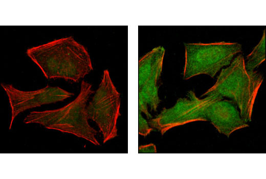
Confocal immunofluorescent analysis of HeLa cells -/+ UV light, labeled with Phospho-p38 MAPK (green). Absence of staining in untreated cells (left) and nuclear localization in treated cells (right). Red = Actin filaments (phalloidin).


