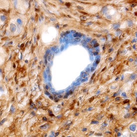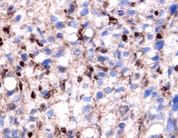
Galectin-1 in Human Prostate Cancer Tissue. Galectin-1 was detected in immersion fixed paraffin-embedded sections of human prostate cancer tissue using 1.7 ug/ml Goat Anti-Human Galectin-1 Antigen Affinity-purified Polyclonal Antibody overnight at 4°C. Tissue was stained with Anti-Goat HRP-DAB (brown) and counterstained with hematoxylin (blue).

Galectin-1 in Human Prostate Cancer Tissue. Galectin-1 was detected in immersion fixed paraffin-embedded sections of human prostate cancer tissue using Goat Anti-Human Galectin-1 Antigen Affinity-purified Polyclonal Antibody at 5 ug/ml overnight at 4°C. Tissue was stained using Anti-Goat HRP-DAB (brown) and counterstained with hematoxylin (blue). Specific labeling was localized to the cytoplasm of stromal cells and nuclei of epithelial cells.

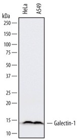
Detection of Human Galectin-1 by Western Blot. Western blot shows lysates of HeLa human cervical epithelial carcinoma cell line and A549 human lung carcinoma cell line. PVDF membrane was probed with 0.1 ug/ml of Goat Anti-Human Galectin-1 Antigen Affinity-purified Polyclonal Antibody followed by HRP-conjugated Anti-Goat IgG Secondary Antibody. A specific band was detected for Galectin-1 at approximately 14 kD (as indicated). This experiment was conducted under reducing conditions.

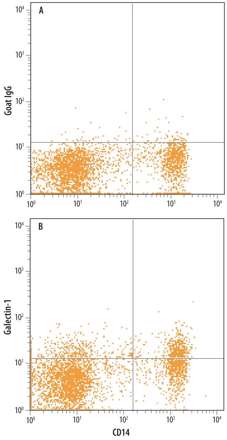
Detection of Galectin-1 in Human Blood Monocytes by Flow Cytometry. Human peripheral blood monocytes were stained with Mouse Anti-Human CD14 PE-conjugated Monoclonal Antibody and either (A) Normal Goat IgG Control or (B) Goat Anti-Human Galectin-1 Antigen Affinity-purified Polyclonal Antibody followed by Allophycocyanin-conjugated Anti-Goat IgG Secondary Antibody. To facilitate intracellular staining, cells were fixed with paraformaldehyde and permeabilized with saponin.


