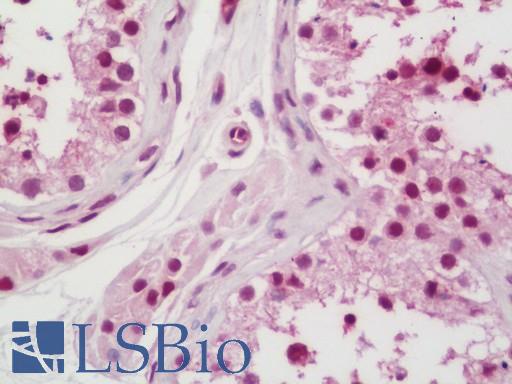
Human Testis: Formalin-Fixed, Paraffin-Embedded (FFPE)

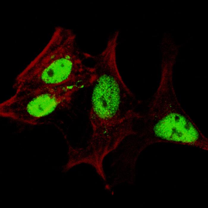
Fluorescent confocal image of SY5Y cells stained LIN28A antibody. SY5Y cells were fixed with 4% PFA (20 min), permeabilized with Triton X-100 (0.2%, 30 min), then incubated LIN28A primary antibody (1:200, 2 h at room temperature). For secondary antibody, Alexa Fluor 488 conjugated donkey anti-rabbit antibody (green) was used (1:1000, 1h). Cytoplasmic actin was counterstained with Alexa Fluor 555 (red) conjugated Phalloidin (5.25 mu M, 25 min). Lin28a immunoreactivity is localized very specifically to the nuclei of the SY5Y cells.

Immunofluorescent of A549 cells, using LIN28A Antibody. Antibody was diluted at 1:100 dilution. Alexa Fluor 488-conjugated goat anti-rabbit lgG at 1:400 dilution was used as the secondary antibody (green). Cytoplasmic actin was counterstained with Dylight Fluor 554 (red) conjugated Phalloidin (red).

LIN28A Antibody western blot of mouse Neuro-2a cell line lysates (35 ug/lane). The LIN28A antibody detected the LIN28A protein (arrow).

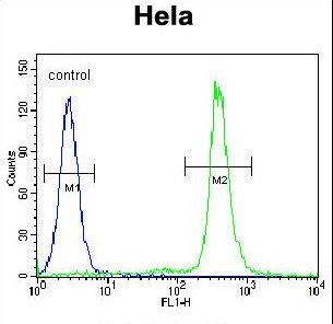
LIN28A Antibody flow cytometry of HeLa cells (right histogram) compared to a negative control cell (left histogram). FITC-conjugated goat-anti-rabbit secondary antibodies were used for the analysis.


