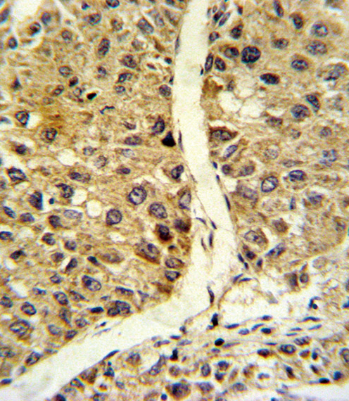
Formalin-fixed and paraffin-embedded human hepatocarcinoma reacted with NOS3 Antibody , which was peroxidase-conjugated to the secondary antibody, followed by DAB staining. This data demonstrates the use of this antibody for immunohistochemistry; clinical relevance has not been evaluated.
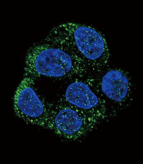
Confocal immunofluorescent of NOS3 Antibody with HepG2 cell followed by Alexa Fluor 488-conjugated goat anti-rabbit lgG (green). DAPI was used to stain the cell nuclear (blue).
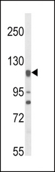
Western blot of NOS3 Antibody in HL-60 cell line lysates (35 ug/lane). NOS3 (arrow) was detected using the purified antibody.
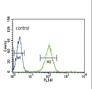
NOS3 Antibody flow cytometry of HL-60 cells (right histogram) compared to a negative control cell (left histogram). FITC-conjugated goat-anti-rabbit secondary antibodies were used for the analysis.