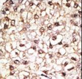
Formalin-fixed and paraffin-embedded human cancer tissue reacted with the primary antibody, which was peroxidase-conjugated to the secondary antibody, followed by AEC staining. This data demonstrates the use of this antibody for immunohistochemistry; clinical relevance has not been evaluated. BC = breast carcinoma; HC = hepatocarcinoma.

Formalin-fixed and paraffin-embedded human testis tissue reacted with PARK2 (Parkin) antibody , which was peroxidase-conjugated to the secondary antibody, followed by DAB staining. This data demonstrates the use of this antibody for immunohistochemistry; clinical relevance has not been evaluated.
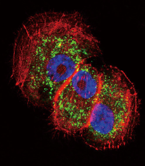
Confocal immunofluorescent of Parkin Antibody with NCI-H460 cell followed by Alexa Fluor 488-conjugated goat anti-rabbit lgG (green). Actin filaments have been labeled with Alexa Fluor 555 phalloidin (red). DAPI was used to stain the cell nuclear (blue).
The anti-Parkin antibody is used in Western blot to detect Parkin in mouse kidney tissue lysate.

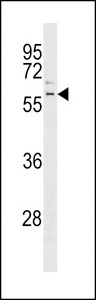
Park2 Antibody western blot of K562 cell line lysates (35 ug/lane). The Park2 antibody detected the Park2 protein (arrow).
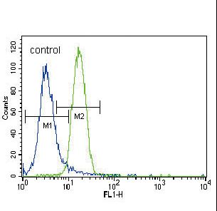
Parkin Antibody flow cytometry of NCI-H460 cells (right histogram) compared to a negative control cell (left histogram). FITC-conjugated goat-anti-rabbit secondary antibodies were used for the analysis.