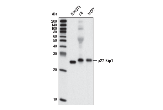
Western blot analysis of extracts from NIH/3T3, C6 and MCF7 cells using p27 Kip1 (SX53G8.5) Mouse mAb.

Confocal immunofluorescent analysis of MCF7 cells using p27 Kip1 (SX53G8.5) Mouse mAb (green). Actin filaments have been labeled with DY-554 phalloidin (red).

Flow cytometric analysis of Jurkat cells using p27 Kip1 (SX53G8.5) Mouse mAb versus Propidium Iodide (PI)/RNase Staining Solution #4087. Anti-mouse IgG (H+L), F(ab') 2 Fragment (Alexa Fluor ® 488 Conjugate) #4408 was used as a secondary Ab.


