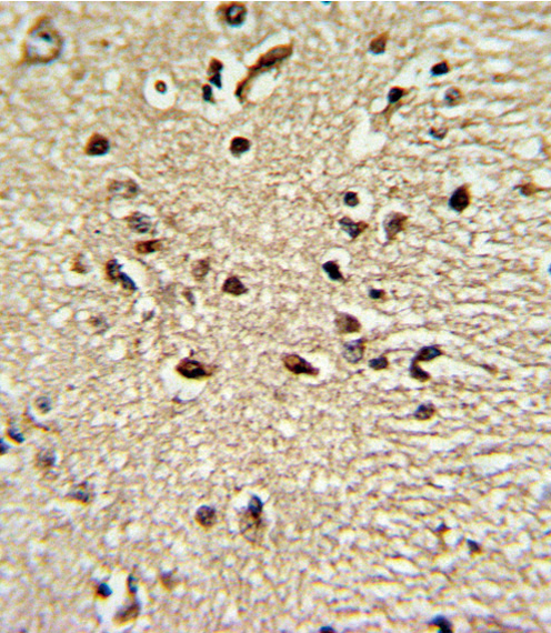
Formalin-fixed and paraffin-embedded human brain tissue reacted with PAX4 Antibody , which was peroxidase-conjugated to the secondary antibody, followed by DAB staining. This data demonstrates the use of this antibody for immunohistochemistry; clinical relevance has not been evaluated.
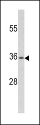
Western blot of PAX4 antibody (RB20216) in CEM cell line lysates (35 ug/lane). PAX4 (arrow) was detected using the purified antibody.
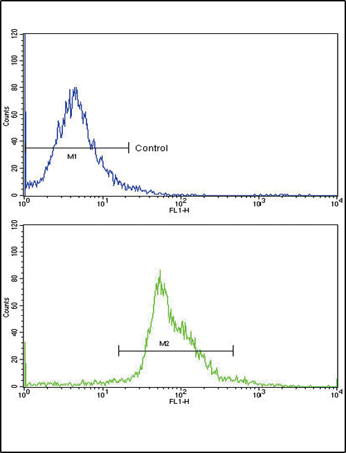
Flow cytometric of widr cells using PAX4 Antibody (bottom histogram) compared to a negative control cell (top histogram)FITC-conjugated goat-anti-rabbit secondary antibodies were used for the analysis.