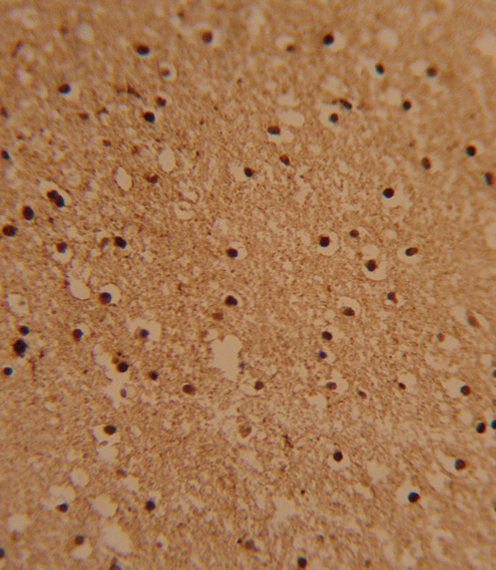
Formalin-fixed and paraffin-embedded human brain tissue reacted with PSMB1 Antibody , which was peroxidase-conjugated to the secondary antibody, followed by DAB staining. This data demonstrates the use of this antibody for immunohistochemistry; clinical relevance has not been evaluated.
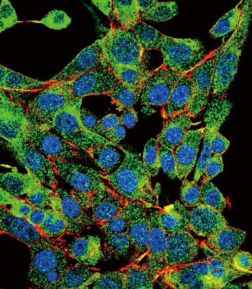
Confocal immunofluorescent of PSMB1 Antibody with HepG2 cell followed by Alexa Fluor 488-conjugated goat anti-rabbit lgG (green). Actin filaments have been labeled with Alexa Fluor 555 phalloidin (red). DAPI was used to stain the cell nuclear (blue).
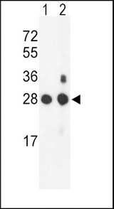
Western blot of PSMB1 Antibody in mouse NIH-3T3 cell line(lane 1) and mouse bladder tissue(lane 2) lysates (35 ug/lane). PSMB1 (arrow) was detected using the purified antibody.
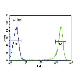
PSMB1 Antibody flow cytometry of HL-60 cells (right histogram) compared to a negative control cell (left histogram). FITC-conjugated goat-anti-rabbit secondary antibodies were used for the analysis.