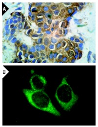
HSP 70 (K-20): sc-1060. Immunoperoxidase staining of formalin-fixed, paraffin-embedded human breast carcinoma tissue (A) and immunofluorescence staining of methanol-fixed HeLa cells (B) showing cytoplasmic localization.
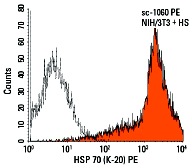
HSP 70 (K-20) PE: sc-1060 PE. Intracellular FCM analysis of fixed and permeabilized, heat-shocked NIH/3T3 cells. Black line histogram represents the isotype control, normal goat IgG: sc-3992.
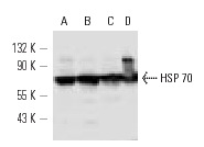
HSP 70 (K-20)-R: sc-1060-R. Western blot analysis of HSP 70 expression in HeLa (A), heat shock-treated HeLa (B), NIH/3T3 (C) and heat shock-treated NIH/3T3 (D) whole cell lysates.
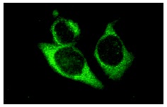
HSP 70 (K-20): sc-1060. Immunofluorescence staining of methanol-fixed HeLa cells showing cytoplasmic localization.
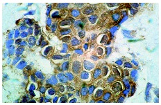
HSP 70 (K-20): sc-1060. Immunoperoxidase staining of formalin-fixed, paraffin-embedded human breast carcinoma tissue showing cytoplasmic localization.
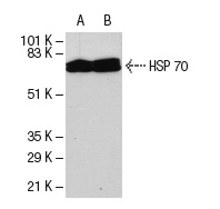
HSP 70 (K-20): sc-1060. Western blot analysis of HSP 70 expression in untreated (A) and heat shock-treated (B) HeLa whole cell lysates.


