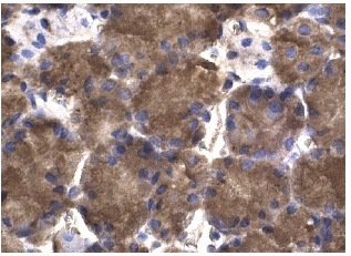
IRS-1 (C-20): sc-559. Immunoperoxidase staining of formalin fixed, paraffin-embedded human tissue showing cytoplasmic staining of glandular cells.
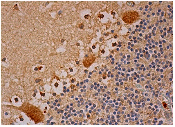
IRS-1 (C-20): sc-559-G. Immunoperoxidase staining of formalin fixed, paraffin-embedded human cerebellum tissue showing cytoplasmic and nuclear staining of Purkinje cells and cytoplasmic staining of cells in granular layer and molecular layer.
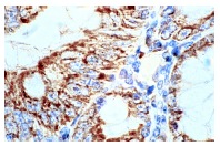
IRS-1 (C-20): sc-559. Immunoperoxidase staining of formalin-fixed, paraffin-embedded human colon carcinoma tissue. Note cytoplasmic staining of mucosal epithelia.

Western blot analysis of IRS-1 expression in 3T3-L1 (A), Ramos (B) and BJAB (C) whole cell lysates. Antibodies tested include IRS-1 (C-20): sc-559 (A,B) and IRS-1 (C-20)-G: sc-559-G (C).
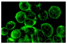
IRS-1 (C-20): sc-559. Immunofluorescence staining of methanol-fixed MCF7 cells showing cytoplasmic localization.
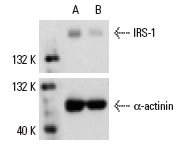
IRS-1 siRNA (h): sc-29376. Western blot analysis of IRS-1 expression in non-transfected control (A) and IRS-1 siRNA transfected (B) HeLa cells. Blot probed with IRS-1 (C-20): sc-559. α-actinin (H-2): sc-17829 used as specificity and loading control.
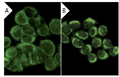
IRS-1 (C-20): sc-559. Immunofluorescence staining of methanol-fixed HeLa cells showing cytoplasmic localization using indirect FITC (A) staining and direct Alexa Fluor 488 (B) staining.
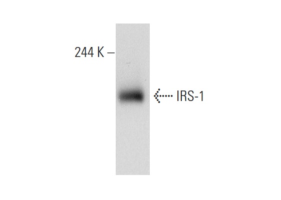
IRS-1 (C-20): sc-559. Western blot analysis of IRS-1 expression in A549 whole cell lysate.
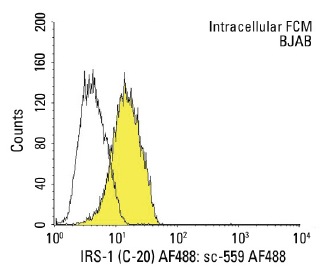
IRS-1 (C-20) AF488: sc-559 AF488. Intracellular FCM analysis of fixed and permeabilized BJAB cells. Black line histogram represents the isotype control, normal rabbit IgG: sc-45068.


