
IRS-1 (A-19): sc-560. Western blot analysis of IRS-1 expression in A549 (A), Ramos (B) and BJAB (C) whole cell lysates.
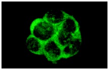
IRS-1 (A-19): sc-560. Immunofluorescence staining of methanol-fixed MCF7 cells showing cytoplasmic localization.
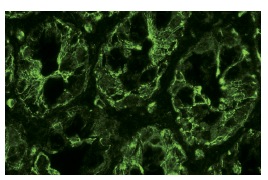
IRS-1 (A-19): sc-560. Immunofluorescence staining of normal mouse intestine frozen section showing cytoplasmic staining.
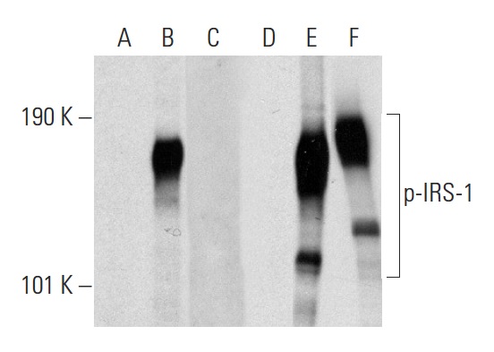
Western blot analysis of IRS-1 phosphorylation in non-transfected: sc-117752 (A,D), untreated human IRS-1 transfected: sc-116569 (B,E) and lambda protein phosphatase (sc-200312A) treated human IRS-1 transfected: sc-116569 (C,F) 293T whole cell lysates. Antibodies tested include p-IRS-1 (Ser 374)-R: sc-17193-R (A,B,C) and IRS-1 (A-19): sc-560 (D,E,F).
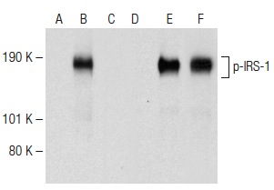
Western blot analysis of IRS-1 phosphorylation in non-transfected: sc-117752 (A,D), untreated human IRS-1 transfected: sc-116569 (B,E) and lambda protein phosphatase (sc-200312A) treated human IRS-1 transfected: sc-116569 (C,F) 293T whole cell lysates. Antibodies tested include p-IRS-1 (Ser 636): sc-33957 (A,B,C) and IRS-1 (A-19): sc-560 (D,E,F).
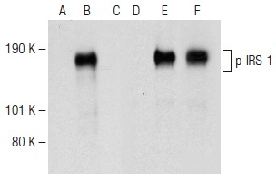
Western blot analysis of IRS-1 phosphorylation in non-transfected: sc-117752 (A,D), untreated human IRS-1 transfected: sc-116569 (B,E) and lambda protein phosphatase (sc-200312A) treated human IRS-1 transfected: sc-116569 (C,F) 293T whole cell lysates. Antibodies tested include p-IRS-1 (Ser 636): sc-101711 (A,B,C) and IRS-1 (A-19): sc-560 (D,E,F).
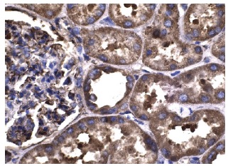
IRS-1 (A-19): sc-560. Immunoperoxidase staining of formalin fixed, paraffin-embedded human kidney tissue showing cytoplasmic staining of cells in glomeruli and tubules.
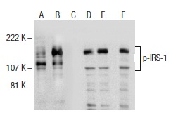
Western blot analysis of IRS-1 phosphorylation in untreated (A,D), insulin treated (B,E) and insulin and lambda protein phosphatase (sc-200312A) treated (C,F) MCF7 whole cell lysates. Antibodies tested include p-IRS-1 (Tyr 465)-R: sc-17194-R (A,B,C) and IRS-1 (A-19): sc-560 (D,E,F).


