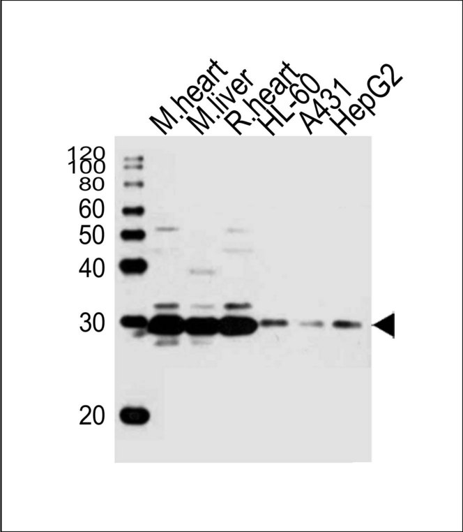
Western blot of lysates from mouse heart, mouse liver, rat heart tissue, HL-60, A431, HepG2 cell line (from left to right) with (DANRE) sdhb Antibody. Antibody was diluted at 1:1000 at each lane. A goat anti-rabbit IgG H&L (HRP) at 1:10000 dilution was used as the secondary antibody.
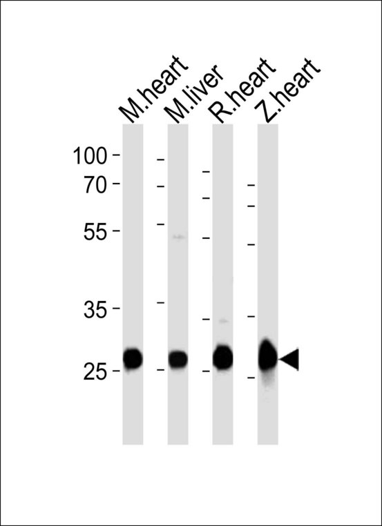
Western blot of lysates from mouse heart, mouse liver, rat heart, zebra fish heart tissue lysate (from left to right) with (DANRE) sdhb Antibody. Antibody was diluted at 1:1000 at each lane. A goat anti-rabbit IgG H&L (HRP) at 1:10000 dilution was used as the secondary antibody.