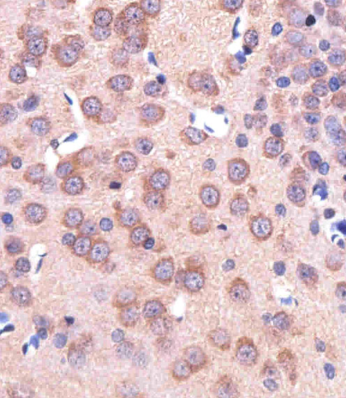
Immunohistochemical of paraffin-embedded R. brain section using ENT1(Slc29a1). Antibody was diluted at 1:25 dilution. A peroxidase-conjugated goat anti-rabbit IgG at 1:400 dilution was used as the secondary antibody, followed by DAB staining.
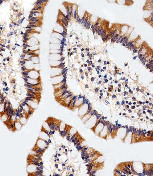
Immunohistochemical of paraffin-embedded H. colon section using ENT1(Slc29a1). Antibody was diluted at 1:25 dilution. A peroxidase-conjugated goat anti-rabbit IgG at 1:400 dilution was used as the secondary antibody, followed by DAB staining.
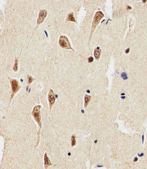
Immunohistochemical of paraffin-embedded H. brain section using ENT1(Slc29a1). Antibody was diluted at 1:25 dilution. A peroxidase-conjugated goat anti-rabbit IgG at 1:400 dilution was used as the secondary antibody, followed by DAB staining.
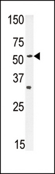
Western blot of ENT1 Antibody in mouse heart tissue lysates (35 ug/lane). ENT1 (arrow) was detected using the purified antibody.

Western blot of lysate from human heart tissue lysate, using ENT1(Slc29a1) Antibody. Antibody was diluted at 1:1000. A goat anti-rabbit IgG H&L (HRP) at 1:5000 dilution was used as the secondary antibody. Lysate at 35ug.