RPA70/RPA1 Antibody
| Name | RPA70/RPA1 Antibody |
|---|---|
| Supplier | Cell Signaling Technology |
| Catalog | 2267 |
| Prices | $246.00 |
| Sizes | 100 µl (10 western blots) |
| Host | Rabbit |
| Clonality | Polyclonal |
| Applications | WB IP ICC/IF FC |
| Species Reactivities | Human, Monkey |
| Antigen | Polyclonal antibodies are produced by immunizing animals with a synthetic peptide corresponding to amino acids near the amino terminus of human RPA70 |
| Description | Rabbit Polyclonal |
| Gene | RPA1 |
| Supplier Page | Shop |
Product images
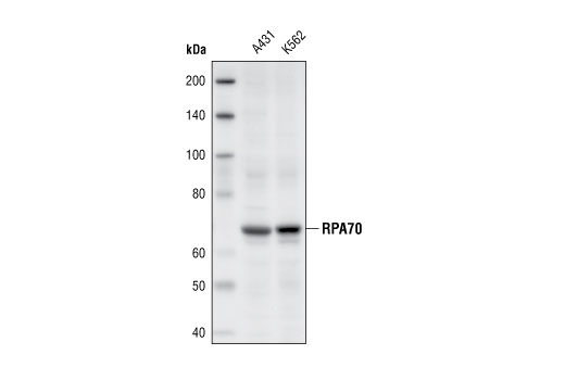 Western blot analysis of extracts from A431 and K562 cells, using RPA70 Antibody.
 Confocal immunofluorescent images of HeLa cells, untreated (left) or UV-treated (right), labeled with RPA70 Antibody (green) showing translocation to distinct nuclear foci after UV damage. Actin filaments have been labeled with Alexa Fluor® 555 phalloidin. Blue pseudocolor = DRAQ5™ (fluorescent DNA dye).
 Flow cytometric analysis of Jurkat cells, using RPA70 antibody (blue) compared to a nonspecific negative control antibody (red).