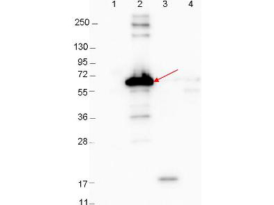
Anti-DbpA Antibody - Western Blot. Western blot showing detection of 0.1 ug recombinant proteins in western blot. Lane 1: Molecular weight markers. Lane 2: MBP-DbpA fusion protein (arrow; expected MW: 60.9 kD). Lane 3: DbpA, MBP removed by TEV cleavage. Lane 4: MBP alone. Protein was run on a 4-20% gel, then transferred to 0.45 micron nitrocellulose. After blocking with 1% BSA-TTBS (MB-013, diluted to 1X) overnight at 4°C, primary antibody was used at 1:1000 at room temperature for 30 min. HRP-conjugated Goat-Anti-Rabbit (p/n LS-C60865) secondary antibody was used at 1:40000 in MB-070 blocking buffer and imaged on the VersaDoc MP 4000 imaging system (Bio-Rad).
