
Recombinant Equine IL‑10 (Catalog # 1605-IL) stimulates proliferation in the MC/9‑2 mouse mast cell line in a dose-dependent manner (orange line). Proliferation elicited by Recombinant Equine IL‑10 (20 ng/mL) is neutralized (green line) by increasing concentrations of Goat Anti-Equine IL‑10 Antigen Affinity-purified Polyclonal Antibody (Catalog # AF1605). The ND50 is typically 0.2-0.6 µg/mL.
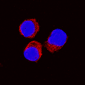
IL‑10 was detected in immersion fixed equine peripheral blood mononuclear cells (PBMCs) treated with calcium ionomycin and PMA using Goat Anti-Equine IL‑10 Antigen Affinity-purified Polyclonal Antibody (Catalog # AF1605) at 15 µg/mL for 3 hours at room temperature. Cells were stained using the NorthernLights™ 557-conjugated Anti-Goat IgG Secondary Antibody (red; Catalog # NL001) and counterstained with DAPI (blue). Specific staining was localized to cytoplasm. View our protocol for Fluorescent ICC Staining of Non-adherent Cells.