
LAMP1/CD107a was detected in immersion fixed RAW 264.7 mouse monocyte/macrophage cell line using Goat Anti-Mouse LAMP1/CD107a Lumenal Domain Antigen Affinity-purified Polyclonal Antibody (Catalog # AF4320) at 10 µg/mL for 3 hours at room temperature. Cells were stained using the NorthernLights™ 557-conjugated Anti-Goat IgG Secondary Antibody (red; Catalog # NL001) and counterstained with DAPI (blue). Specific staining was localized to cytoplasm. View our protocol for Fluorescent ICC Staining of Cells on Coverslips.
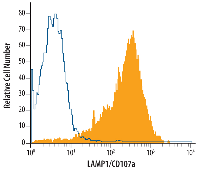
RAW 264.7 mouse monocyte/macrophage cell line was stained with Goat Anti-Mouse LAMP1/CD107a Lumenal Domain Antigen Affinity-purified Polyclonal Antibody (Catalog # AF4320, filled histogram) or control antibody (Catalog # AB-108-C, open histogram), followed by Allophycocyanin-conjugated Anti-Goat IgG Secondary Antibody (Catalog # F0108). To facilitate intracellular staining, cells were fixed with Flow Cytometry Fixation Buffer (Catalog # FC004) and permeabilized with Flow Cytometry Permeabilization/Wash Buffer I (Catalog # FC005).View our protocol for Staining Intracellular Molecules.

Western blot shows lysates of mouse small intestine tissue and NIH‑3T3 mouse embryonic fibroblast cell line. PVDF membrane was probed with 0.2 µg/mL of Goat Anti-Mouse LAMP‑1/CD107a Lumenal Domain Antigen Affinity-purified Polyclonal Antibody (Catalog # AF4320) followed by HRP-conjugated Anti-Goat IgG Secondary Antibody (Catalog # HAF017). A specific band was detected for LAMP‑1/CD107a at approximately 100-120 kDa (as indicated). This experiment was conducted under reducing conditions and using Immunoblot Buffer Group 1.