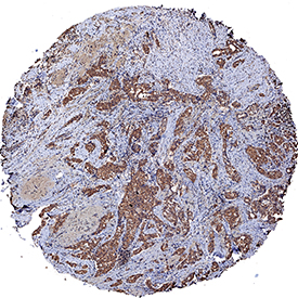
TM4SF1/L6 was detected in immersion fixed paraffin-embedded sections of human breast cancer tissue using Mouse Anti-Human TM4SF1/L6 Monoclonal Antibody (Catalog # MAB8164) at 5 µg/mL for 1 hour at room temperature followed by incubation with the Anti-Mouse IgG VisUCyte™ HRP Polymer Antibody (Catalog # VC001). Tissue was stained using DAB (brown) and counterstained with hematoxylin (blue). Specific staining was localized to cytoplasm in cancer cells. View our protocol for IHC Staining with VisUCyte HRP Polymer Detection Reagents.

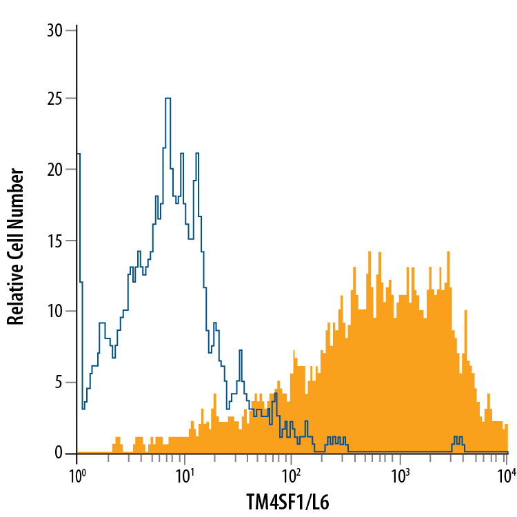
A549 human lung carcinoma cell line was stained with Mouse Anti-Human TM4SF1/L6 Monoclonal Antibody (Catalog # MAB8164, filled histogram) or isotype control antibody (Catalog # MAB002, open histogram), followed by Allophycocyanin-conjugated Anti-Mouse IgG Secondary Antibody (Catalog # F0101B).

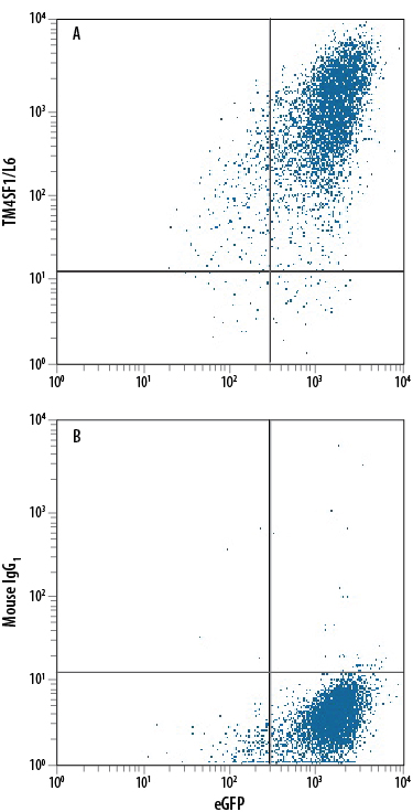
HEK293 human embryonic kidney cell line transfected with human TM4SF1/L6 and eGFP was stained with either (A) Mouse Anti-Human TM4SF1/L6 Monoclonal Antibody (Catalog # MAB8164) or (B) Mouse IgG1 Isotype Control (Catalog # MAB002) followed by Phycoerythrin-conjugated Anti-Mouse IgG Secondary Antibody (Catalog # F0102B).

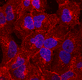
TM4SF1/L6 was detected in immersion fixed A549 human lung carcinoma cell line using Mouse Anti-Human TM4SF1/L6 Monoclonal Antibody (Catalog # MAB8164) at 10 µg/mL for 3 hours at room temperature. Cells were stained using the NorthernLights™ 557-conjugated Anti-Mouse IgG Secondary Antibody (red; Catalog # NL007) and counterstained with DAPI (blue). Specific staining was localized to cell membranes. View our protocol for Fluorescent ICC Staining of Cells on Coverslips.


