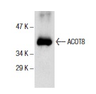
ACOT8 (H-320): sc-7018. Western blot analysis of human recombinant ACOT8.
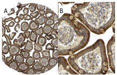
TEII p35 (H-320): sc-7018. Immunoperoxidase staining of formalin fixed, paraffin-embedded human small intestine tissue showing cytoplasmic staining of glandular cells at low (A) and high (B) magnification. Kindly provided by The Swedish Human Protein Atlas (HPA) program.
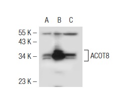
ACOT8 (H-320): sc-7018. Western blot analysis of ACOT8 expression in non-transfected 293T: sc-117752 (A), mouse ACOT8 transfected 293T: sc-126376 (B) and Ramos (C) whole cell lysates.