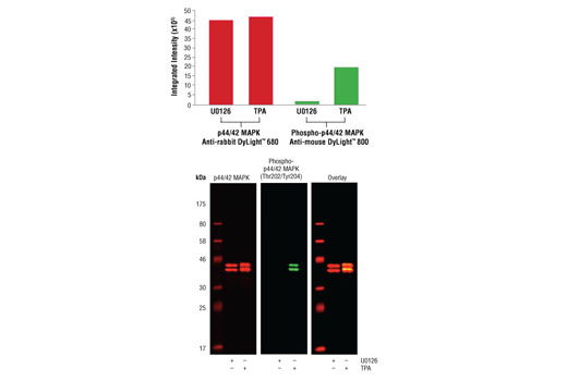
Western blot analysis of Jurkat cell lysates (#9194) treated with either U0126 (MEK 1/2 inhibitor) #9903 or TPA (12-O-Tetradecanoylphorbol-13-Acetate) #4174, using Phospho-p44/42 MAPK (Erk1/2) (Thr202/204) (D13.14.4E) XP ® Rabbit mAb #4370 detected with Anti-rabbit IgG (H+L) (DyLight™ 800 Conjugate) #5151 (green) and p44/42 MAPK (Erk1/2) (3A7) Mouse mAb #9107 detected with Anti-mouse IgG (H+L) (DyLight™ 680 Conjugate) (red). The array image pixel intensities obtained using a LI-COR ® Biosciences Odyssey ® Infrared Imaging System are shown in the upper panel while corresponding fluorescent western blots are shown in the lower panel.

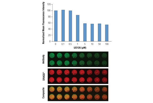
In-Cell Western™ analysis of A549 cells exposed to varying concentrations of U0126 (MEK1/2 Inhibitor) #9903 for 3 hours, followed by TPA (Phorbol-12-Myristate-13-Acetate) #9905 stimulation for 30 minutes. With increasing concentrations of U0126, a significant decrease (~2 fold) in Phospho-p44/42 MAPK (Erk1/2) (Thr202/Tyr204) (E10) Mouse mAb #9106 signal as compared to the TPA-stimulated control was observed. Data and images were generated on the LI-COR ® Biosciences Odyssey ® Infrared Imaging System using Anti-mouse IgG (H+L) (DyLight™ 800 4X PEG Conjugate). DRAQ5 ® #4084 (fluorescent DNA dye - red) is used for normalization.

