![Western blot analysis of extracts from various cell lines and primary cell cultures, using VEGF Receptor 2 (55B11) Rabbit mAb. PAE/CKR cells overexpress chimeric receptors containing human CSF-1 extracellular binding domain/mouse VEGF receptor 2 intracellular domains (Rahimi, N. et al. [2000] J. Biol. Chem. 275, 16986-16992). PAE/VEGFR1 cells overexpress human VEGF receptor 1.](http://www.bioprodhub.com/system/product_images/ab_products/1/sub_2/605_2479_wb_jb_040714.jpg)
Western blot analysis of extracts from various cell lines and primary cell cultures, using VEGF Receptor 2 (55B11) Rabbit mAb. PAE/CKR cells overexpress chimeric receptors containing human CSF-1 extracellular binding domain/mouse VEGF receptor 2 intracellular domains (Rahimi, N. et al. [2000] J. Biol. Chem. 275, 16986-16992). PAE/VEGFR1 cells overexpress human VEGF receptor 1.

Immunohistochemical analysis of paraffin-embedded human astrocytoma, using VEGF Receptor 2 (55B11) Rabbit mAb.

Immunohistochemical analysis of paraffin-embedded breast angiosarcoma, using VEGF Receptor 2 (55B11) Rabbit mAb (left). A serial section is stained for CD31 (PECAM-1), an endothelial cell marker (right).

Immunohistochemical analysis of paraffin-embedded HUVEC cells, using VEGF Receptor 2 (55B11) Rabbit mAb.
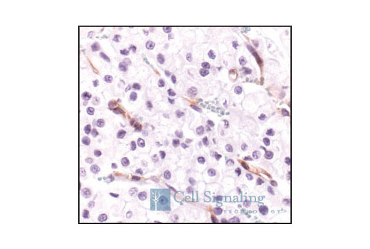
Immunohistochemical analysis of paraffin-embedded human renal adenocarcinoma, using VEGF Receptor 2 (55B11) Rabbit mAb.

Immunohistochemical analysis of paraffin-embedded HT-29 xenograft, using VEGF Receptor 2 (55B11) Rabbit mAb. Note staining of mouse blood vessels.

Confocal immunofluorescent analysis of mouse pancreas using VEGF Receptor 2 (55B11) Rabbit mAb (green) and S6 Ribosomal Protein (54D2) Mouse mAb #2317 (red). Blue pseudocolor = DRAQ5 ® #4084 (fluorescent DNA dye).

Confocal immunofluorescent images of HUVEC cells untreated (left) or stimulated with Vascular Endothelial Growth Factor (VEGF) #9943 (right) and labeled with Phospho-VEGF Receptor 2 (Tyr1175) (19A10) Rabbit mAb #2478 (top, green) and VEGF Receptor 2 (55B11) Rabbit mAb (bottom, green). Blue pseudocolor = DRAQ5 ® (fluorescent DNA dye).