Atg5 (D6M7Q) Rabbit mAb
| Name | Atg5 (D6M7Q) Rabbit mAb |
|---|---|
| Supplier | Cell Signaling Technology |
| Catalog | 13007 |
| Host | Rabbit |
| Clonality | Monoclonal |
| Clone | D6M7Q |
| Applications | WB IP ICC/IF |
| Species Reactivities | Rabbit, Monkey |
| Antigen | Monoclonal antibody is produced by immunizing animals with a synthetic peptide corresponding to residues surrounding Gly232 of human Atg5 protein. |
| Description | Rabbit Monoclonal |
| Gene | ATG5 |
| Supplier Page | Shop |
Product images
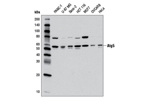 Western blot analysis of extracts from various cell lines using Atg5 (D6M7Q) Rabbit mAb.
 Immunoprecipitation of Atg5 from PANC-1 cell extracts using Rabbit (DA1E) mAb IgG XP® Isotype Control #3900 (lane 2) or Atg5 (D6M7Q) Rabbit mAb (lane 3). Lane 1 is 10% input. Western blot was performed using Atg5 (D6M7Q) Rabbit mAb. Mouse Anti-rabbit IgG (Conformation Specific) (L27A9) mAb #3678 was used as a secondary antibody to avoid cross-reactivity with rabbit IgG.
 Confocal immunofluorescent analysis of HCT 116 cells, untreated (left), nutrient starved with EBSS (4 hr, middle), or chloroquine-treated (50 μM, overnight; right) using Atg5 (D6M7Q) Rabbit mAb (green). Blue pseudocolor = DRAQ5® #4084 (fluorescent DNA dye).