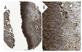
PLC δ1 (H-140): sc-30062. Immunoperoxidase staining of formalin fixed, paraffin-embedded human urinary bladder tissue showing membrane and cytoplasmic staining of surface epithelial cells at low (A) and high (B) magnification. Kindly provided by The Swedish Human Protein Atlas (HPA) program.
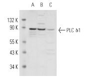
PLC δ1 (H-140): sc-30062. Western blot analysis of PLC δ1 expression in A-10 (A), F9 (B) and NTERA-2 cl.D1 (C) whole cell lysates.
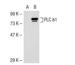
PLC δ1 (H-140): sc-30062. Western blot analysis of PLCδ1 expression in non-transfected: sc-117752 (A) and mouse PLCδ1 transfected: sc-122626 (B) 293T whole cell lysates.