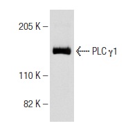
PLC γ1 (E-12): sc-7290. Western blot analysis of PLC γ1 expression in A-431 whole cell lysate.
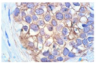
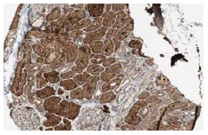
PLC γ1 (E-12): sc-7290. Immunoperoxidase staining of formalin-fixed, paraffin-embedded human colon carcinoma tissue showing cytoplasmic and membrane localization.
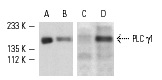
Western blot detection of PLC γ1 phosphorylation in untreated (A,C) and PMA-treated (B,D) NIH/3T3 whole cell lysates. Blots were probed with PLC γ1 (E-12): sc-7290 (A,B) and p-PLC γ1 (Tyr 783)-R: sc-12943-R (C,D).
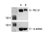
PLC γ1 siRNA (h): sc-29452. Western blot analysis of PLC γ1 expression in non-transfected control (A) and PLC γ1 siRNA transfected (B) HeLa cells. Blot probed with PLC γ1 (E-12): sc-7290. α-actinin (H-2): sc-17829 used as specificity and loading control.
PLC γ1 (E-12): sc-7290. Immunoperoxidase staining of formalin fixed, paraffin-embedded human kidney tissue showing cytoplasmic staining of cells in glomeruli and tubuli. Kindly provided by The Swedish Human Protein Atlas (HPA) program.
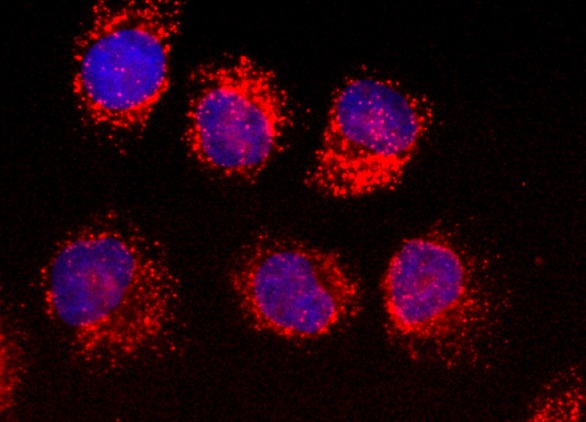
PLC γ1 (E-12): sc-7290. Direct immunofluorescence staining of formalin-fixed HeLa cells showing membrane ruffle localization and nuclear DAPI counterstain. PLC γ1 (E-12) antibody was conjugated to CruzFluor™ 594 succinimidyl ester: sc-362619.
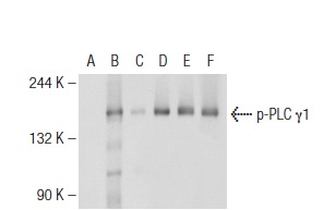
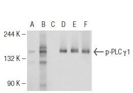
Western blot analysis of PLC γ1 phosphorylation in untreated (A,D), pervanadate treated (B,E) and pervanadate and lambda protein phosphatase treated (C,F) Jurkat whole cell lysates. Antibodies tested include p-PLC γ1 (Tyr 783)-R: sc-12943-R (A,B,C) and PLC γ1 (E-12): sc-7290 (D,E,F).
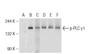
Western blot analysis of PLC γ1 phosphorylation in untreated (A,D), pervanadate treated (B,E) and pervanadate and lambda protein phosphatase treated (C,F) Jurkat whole cell lysates. Antibodies tested include p-PLC γ1 (pY783.27): sc-136186 (A,B,C) and PLC γ1 (E-12): sc-7290 (D,E,F).
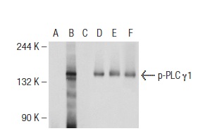
Western blot analysis of PLC γ1 phosphorylation in untreated (A,D), pervanadate treated (B,E) and pervanadate and lambda protein phosphatase treated (C,F) Jurkat whole cell lysates. Antibodies tested include p-PLC γ1 (Tyr 1253)-R: sc-12944-R (A,B,C) and PLC γ1 (E-12): sc-7290 (D,E,F).
Western blot analysis of PLC γ1 phosphorylation in untreated (A,D), pervanadate treated (B,E) and pervanadate and lambda protein phosphatase treated (C,F) Jurkat whole cell lysates. Antibodies tested include p-PLC γ1 (Tyr 1253)-R: sc-22141-R (A,B,C) and PLC γ1 (E-12): sc-7290 (D,E,F).
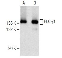
PLC γ1 (E-12): sc-7290. Western blot analysis of PLC γ1 expression in MDBK (A) and MCF7 (B) whole cell lysates.
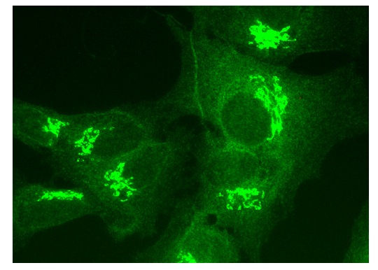
PLC γ1 (E-12): sc-7290. Immunofluorescence staining of formalin-fixed HepG2 cells showing ruffle localization.
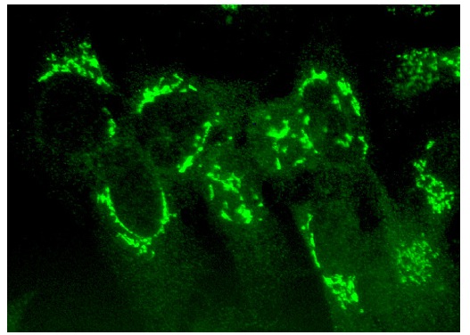
PLC γ1 (E-12): sc-7290. Immunofluorescence staining of formalin-fixed HepG2 cells showing membrane ruffle localization.