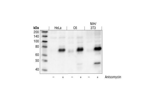
Western blot analysis of extracts from HeLa, C6 and NIH/3T3 cells, untreated or anisomycin-treated, using Phospho-ATF-2 (Thr71) Antibody.

Western blot analysis of extracts from 293 and NIH/3T3 cells, untreated or UV-treated, using ATF-2 (20F1) Rabbit mAb.

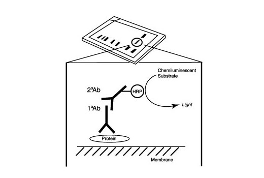
After the primary antibody is bound to the target protein, a complex with HRP-linked secondary antibody is formed. The LumiGLO ® is added and emits light during enzyme catalyzed decomposition.

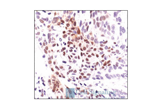
Immunohistochemical analysis of paraffin-embedded human breast carcinoma showing nuclear localization, using Phospho-ATF-2 (Thr71) Antibody .

Immunohistochemical analysis of paraffin-embedded human endometrial carcinoma using ATF-2 (20F1) Rabbit mAb.

Immunohistchemical analysis of paraffin-embedded human colon carcinoma using Phospho-ATF-2 (Thr71) Antibody.

Immunohistochemical analysis of paraffin-embedded human lung carcinoma using Phospho-ATF-2 (Thr71) Antibody.

Immunhistochemical analysis of frozen H1650 xenograft using Phospho-ATF-2 (Thr71) Antibody.

Flow cytometric analysis of Jurkat cells, untreated (blue) or Anisomycin-treated (green), using Phospho-ATF-2 (Thr71) Antibody compared to a nonspecific negative control antibody (red).

Confocal immunofluorescent analysis of HeLa cells, either untreated (left) or anisomycin-treated (right), using Phospho-ATF-2 (Thr71) Antibody (green). Actin filaments have been labeled with Alexa Fluor® 555 phalloidin (red).


