 Immunofluorescence
Nuclear localization of HOXA2 in NIH-3T3 cells over-expressing human HOXA2. Cells were fixed and stained with Rabbit Anti-HOXA2 (1:1,000) (
Cat. No. H9665) followed by Goat Anti-Rabbit, Cy3 conjugate and counterstained with DAPI (blue) to stain nuclei (x40 magnification).
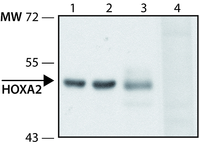
 Immunoblotting
Lysates of the indicated sources were separated on SDS-PAGE and probed with Rabbit Anti-HOXA2 (
Cat. No. H9665) at 1:500 dilution. The antibody was developed with Goat Anti-Rabbit IgG, Peroxidase conjugate (
Cat. No. A0545) and a chemiluminescent substrate.
Lanes1. Mouse lung
2. Mouse liver
3. 293-T over-expressing human HOXA2
4. 293-T over-expressing human HOXA2 + 10 μg/mL HOXA2 immunizing peptide

