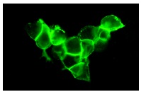
NCAM (ERIC 1): sc-106. Immunofluorescence staining of methanol-fixed SK-N-SH cells showing membrane staining.
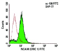
NCAM (ERIC 1) FITC: sc-106 FITC. FCM analysis of SHP-77 cells. Black line histogram represents the isotype control, normal mouse IgG
1: sc-2855.
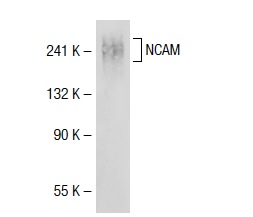
NCAM (ERIC 1): sc-106. Western blot analysis of NCAM expression in IMR-32 whole cell lysate.
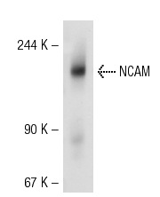
NCAM (ERIC 1): sc-106. Western blot analysis of NCAM expression in 293T whole cell lysate.
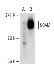
NCAM (ERIC 1): sc-106. Western blot analysis of NCAM expression in non-transfected: sc-117752 (A) and human NCAM transfected: sc-115782 (B) 293T whole cell lysates.
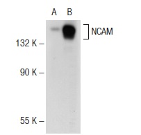
NCAM (ERIC 1): sc-106. Western blot analysis of NCAM expression in non-transfected: sc-117752 (A) and human NCAM transfected: sc-177603 (B) 293T whole cell lysates.
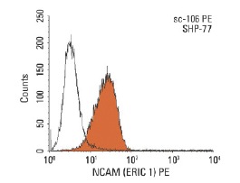
NCAM (ERIC 1) PE: sc-106 PE. FCM analysis of SHP-77 cells. Black line histogram represents the isotype control, normal mouse IgG
1: sc-2866.
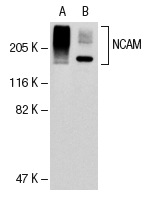
NCAM (ERIC 1): sc-106. Western blot analysis of NCAM expression in IMR-32 (A) and SK-N-SH (B) whole cell lysates.
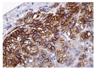
NCAM (ERIC 1): sc-106. Immunoperoxidase staining of formalin fixed, paraffin-embedded human adrenal gland tissue showing membrane staining of subset of glandular cells.


