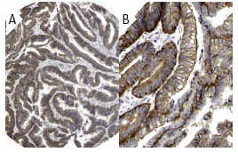
Adducin α (H-100): sc-25731. Immunoperoxidase staining of formalin fixed, paraffin-embedded human colo-rectal cancer tissue showing membrane and cytoplasmic staining of tumor cells at low (A) and high (B) magnification. Kindly provided by The Swedish Human Protein Atlas (HPA) program.
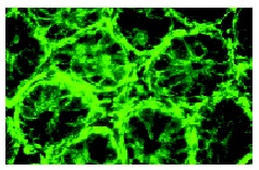
Adducin α (H-100): sc-25731. Immunofluorescence staining of normal mouse intestine frozen section showing membrane and cytoskeletal staining.
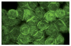
Adducin α (H-100): sc-25731. Immunofluorescence staining of methanol-fixed HeLa cells showing membrane localization.
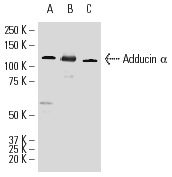
Adducin α (H-100): sc-25731. Western blot analysis of Adducin α expression in SK-N-MC (A) and T98G (B) whole cell lysates and rat spleen tissue extract (C).
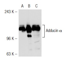
Adducin α (H-100): sc-25731. Western blot analysis of Adducin α expression in non-transfected 293T: sc-117752 (A), mouse Adducin α transfected 293T: sc-118250 (B) and T98G (C) whole cell lysates.
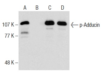
Western blot analysis of Adducin phosphorylation in untreated (A,C) and lambda protein phosphatase (sc-200312A) treated (B,D) HL-60 whole cell lysates. Antibodies tested include p-Adducin (Ser 662)-R: sc-12614-R (A,B) and Adducin α (H-100): sc-25731 (C,D).
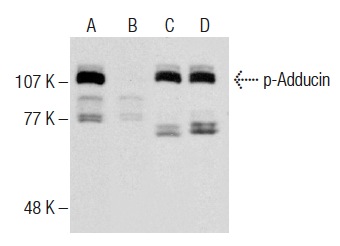
Western blot analysis of Adducin phosphorylation in untreated (A,C) and lambda protein phosphatase (sc-200312A) treated (B,D) rat brain tissue extracts. Antibodies tested include p-Adducin (Ser 662)-R: sc-12614-R (A,B) and Adducin α (H-100): sc-25731 (C,D).
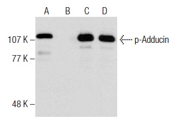
Western blot analysis of Adducin phosphorylation in untreated (A,C) and lambda protein phosphatase (sc-200312A) treated (B,D) HL-60 whole cell lysates. Antibodies tested include p-Adducin (Ser 726)-R: sc-16736-R (A,B) and Adducin α (H-100): sc-25731 (C,D).
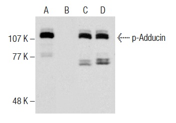
Western blot analysis of Adducin phosphorylation in untreated (A,C) and lambda protein phosphatase (sc-200312A) treated (B,D) rat brain tissue extracts. Antibodies tested include p-Adducin (Ser 726)-R: sc-16736-R (A,B) and Adducin α (H-100): sc-25731 (C,D).


