Anti-ATP6V1C1 (21-35) antibody produced in rabbit
| Name | Anti-ATP6V1C1 (21-35) antibody produced in rabbit |
|---|---|
| Supplier | Sigma-Aldrich |
| Catalog | A1111 |
| Prices | $328.50 |
| Sizes | 200 µl |
| Host | Rabbit |
| Clonality | Polyclonal |
| Applications | WB |
| Species Reactivities | Human |
| Antigen | synthetic peptide corresponding to amino acids 21-35 of human ATP6V1C1 |
| Description | Rabbit Polyclonal |
| Gene | ATP6V1C1 |
| Conjugate | unconjugated |
| Supplier Page | Shop |
Product images
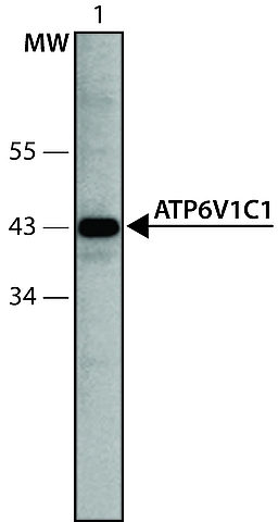 Immunoblotting
Lysate of HEK-293T cells overexpressing human ATP6V1C1 was separated on SDS-PAGE, blotted with Anti-ATP6V1C1 (21-35) (Cat. No. A1111) and developed using Goat Anti-Rabbit IgG-Peroxidase (Cat. No. A0545) and a chemiluminescent substrate.
Lanes
1. Antibody dilution 1:250
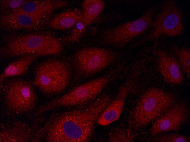 Immunofluorescence
Anti-ATP6V1C1 (21-35): Cat. No. A1111: Immunofluorescence of HUVEC cells using ATP6V1C1 (21-35), Cat. No. A1111 (red) at a 1:100 dilution, taken at 40× magnification and nuclear staining with Hoescht 33342 (blue).Yale HTCB IF procedure used. Images may have been adjusted to improve viewing quality. If you would like the original image, please send a request to protocols@sial.com
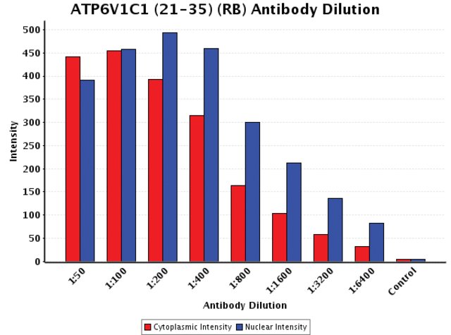 Chart
ChartAnti-ATP6V1C1 (21-35): Cat. No. A1111: Intensity analysis of Anti-ATP6V1C1 (21-35) staining. Cytoplasmic and nuclear intensity values were obtained at 8 different antibody dilutions (1:50 to 1:6,400) as compared with the negative control. Yale HTCB IF procedure used.