 Immunofluorescence
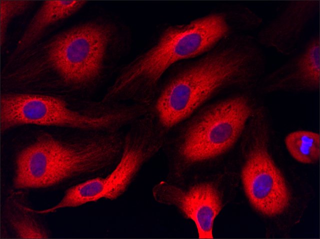
Anti-RANBP1 (6-20),
Cat. No. SAB1100310: Immunofluorescence of HUVEC cells using Anti-RANBP1 (6-20), SAB1100310 (red), taken at 40× magnification and nuclear staining with Hoescht 33342 (blue). Yale HTCB IF procedure used. Images may have been adjusted to improve viewing quality. If you would like the original image, please send a request to protocols@sial.com
 Immunoblotting
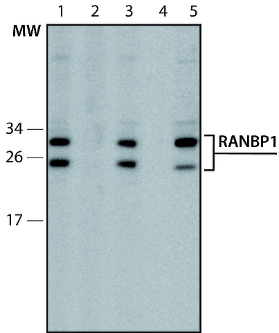
Lysate of HEK-293T cells overexpressing human FLAG-RANBP1 fusion protein was separated on SDS-PAGE, blotted with Anti-RANBP1 (6-20) (
Cat. No. SAB1100310) and developed using Goat Anti-Rabbit IgG-Peroxidase (
Cat. No. A0545) and a chemiluminescent substrate.
Lanes1. Antibody dilution 1:500
2. Antibody dilution 1:500 + RANBP1 immunizing peptide (human, 6-20)
3. Antibody dilution 1:1,000
4. Negative control (only secondary antibody)
5. Anti-FLAG antibody (
Cat. No. F7425), working concentration 2 μg/mL
 Immunofluorescence Graph
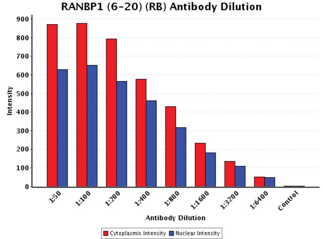
Anti-RANBP1 (6-20),
Cat. No. SAB1100310: Intensity analysis of Anti-RANBP1 (6-20) staining. Cytoplasmic and nuclear intensity values were obtained at 8 different Antibody dilutions (1:50 to 1:6,400) as compared with the negative control. Yale HTCB IF procedure used.


