
FAS (B-10): sc-8009. Western blot analysis of FAS expression in A-431 whole cell lysate.
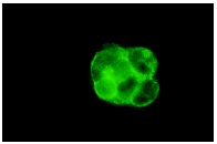
FAS (B-10): sc-8009. Immunofluorescence staining of methanol-fixed MCF7 cells showing membrane localization.
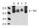
FAS (B-10): sc-8009. Western blot analysis of FAS expression in A-431 (A), MDA-MB-468 (B), Jurkat (C) and Caki-1 (D) whole cell lysates.
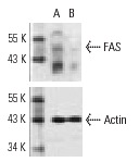
FAS siRNA (h): sc-29311. Western blot analysis of FAS expression in non-transfected control (A) and FAS siRNA transfected (B) HeLa cells. Blot probed with FAS (B-10): sc-8009. Actin (I-19): sc-1616 used as specificity and loading control.
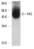
Western blot analysis of FAS expression in A-431 whole cell lysate immunoprecipitated with FAS (C236): sc-21730 and detected with FAS (B-10): sc-8009.
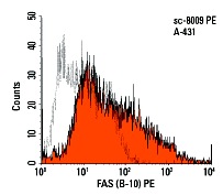
FAS (B-10) PE: sc-8009 PE. FCM analysis of A-431 cells. Black line histogram represents the isotype control, normal mouse IgG
1: sc-2866.
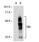
FAS (B-10): sc-8009. Western blot analysis of FAS expression in non-transfected: sc-117752 (A) and human FAS transfected: sc-113770 (B) 293T whole cell lysates.
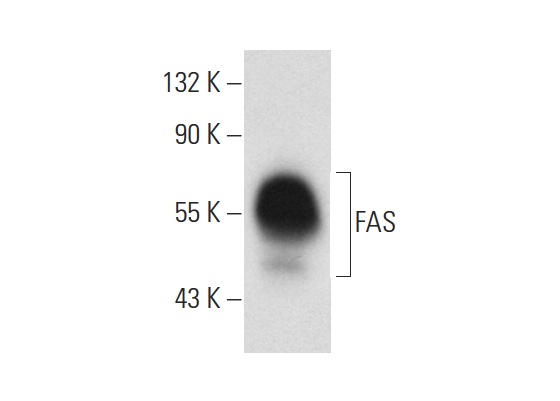
FAS (B-10): sc-8009. Western blot analysis of FAS expression in HuT 78 whole cell lysate.
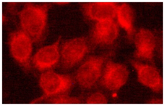
FAS (B-10): sc-8009. Immunofluorescence staining of methanol-fixed HeLa cells showing membrane localization.
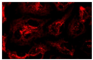
FAS (B-10): sc-8009. Immunofluorescence staining of methanol-fixed HeLa cells showing membrane localization.
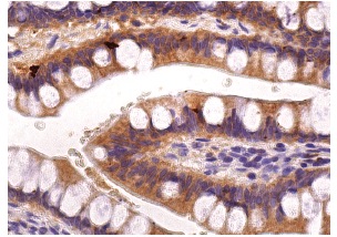
FAS (B-10): sc-8009. Immunoperoxidase staining of formalin fixed, paraffin-embedded human small intestine tissue showing cytoplasmic staining of glandular cells.
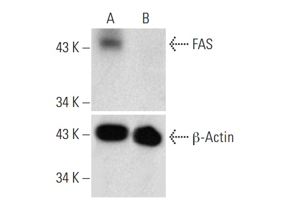
FAS HDR Plasmid (h): sc-400481-HDR. Western blot analysis of FAS expression in non-transfected control(A) and puromycin treated, FAS CRISPR/Cas9 KO Plasmid: sc-400481 plus FAS HDR Plasmid co-transfected (B) HEK 293T whole cell lysates. Blot probed with FAS (B-109): sc-8009. β-Actin (H-2): sc-47778 used as specificity and loading control.


