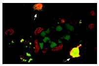
FAS-L (Q-20): sc-956. FAS-L expression in MS plaque. Confocal microscopy showing cells labeled by TUNEL (yellow) and sc-956 (red). Arrows indicate double-labeled cells. Reproduced from P. Dowling et al.,The Journal of Experimental Medicine, 1996, vol: 184, 1513-1518 by copyright permission of the Rockefeller University Press.
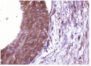
FAS-L (Q-20): sc-956. Immunoperoxidase staining of formalin fixed, paraffin-embedded human testes tissue showing cytoplasmic staining of cells in seminiferus ducts.
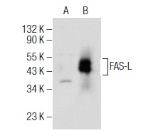
FAS-L (Q-20): sc-956. Western blot analysis of FAS-L expression in non-transfected: sc-117752 (A) and human FAS-L transfected: sc-159339 (B) 293T whole cell lysates.
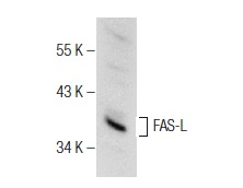
FAS-L (Q-20): sc-956. Western blot analysis of FAS-L expression in Jurkat whole cell lysate.
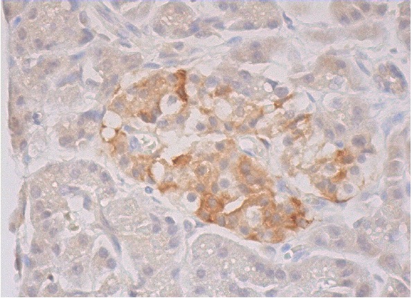
FAS-L (Q-20)-G: sc-956-G. Immunoperoxidase staining of formalin fixed, paraffin-embedded human pancreas tissue showing cytoplasmic staining of exocrine glandular cells and cytoplasmic and membrane staining of Islets of Langerhans.


