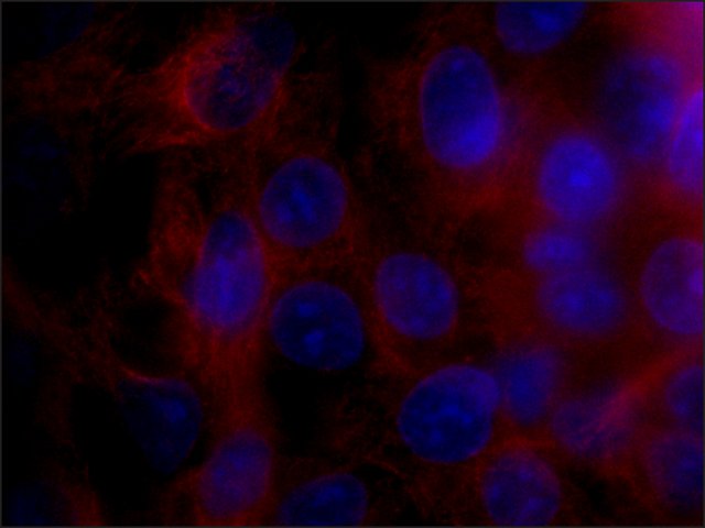
 Immunofluorescence
HepG2 cells were fixed and permeabilized with cold methanol followed by aceton. Fixed cells were stained with Mouse Anti-TUBA8 Clone: TUBA8-C14 (
Cat. No. SAB4100010) diluted to 1:10. The antibody was developed using Goat Anti-Mouse IgG, Cy3 conjugate.
 Immunoblotting
Lysate of HEK-293T cells expressing FLAG
®-tagged human TUBA8 was separated on SDS-PAGE, probed with Mouse Anti-TUBA8 Clone: TUBA8-C14 (
Cat. No. SAB4100010). The antibody was developed using Goat Anti-Mouse IgG-Peroxidase (
Cat. No. A2304) and a chemiluminescent substrate.
Lanes1. Antibody dilution 1:500
2. Antibody dilution 1:1,000
3. Negative control
4. Positive control Anti-FLAG
®
 Immunoblotting
HepG2 cell extract was separated on SDS-PAGE and probed with Mouse Anti-TUBA8 Clone: TUBA8-C14 (
Cat. No. SAB4100010). The antibody was developed using Goat Anti-Mouse IgG-Peroxidase (
Cat. No. A2304) and a chemiluminescent substrate.
Lanes1. Antibody dilution 1:250
2. Antibody dilution 1:500
3. Negative control
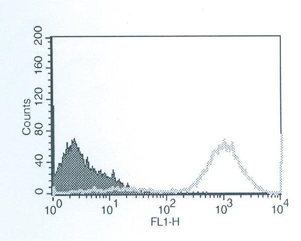
 Flow Cytometry
1×10
6 HT29 cells were fixed and permeabilized with 2% Paraformaldehyde and 0.1% Triton X-100. Fixed cells were labeled with Mouse Anti-TUBA8 Clone: TUBA8-C14 (
Cat. No. SAB4100010) diluted to 1:10 followed by Rabbit Anti-Mouse IgG, FITC conjugate. Shaded histogram: negative control without first antibody, Gray histogram: antibody specific staining.


