Phospho-53BP1 (Ser1778) Antibody
| Name | Phospho-53BP1 (Ser1778) Antibody |
|---|---|
| Supplier | Cell Signaling Technology |
| Catalog | 2675 |
| Prices | $287.00 |
| Sizes | 100 µl (10 western blots) |
| Host | Rabbit |
| Clonality | Polyclonal |
| Applications | WB ICC/IF FC |
| Species Reactivities | Human, Monkey |
| Antigen | Polyclonal antibodies are produced by immunizing animals with a synthetic phosphopeptide corresponding to residues surrounding Ser1778 of human 53BP1 |
| Description | Rabbit Polyclonal |
| Gene | TP53BP1 |
| Supplier Page | Shop |
Product images
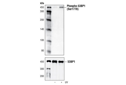 Western blot analysis of extracts from 293 cells, untreated or UV-treated (50 mJ for 2 hours), using Phospho-53BP1 (Ser1778) Antibody (upper) or 53BP1 Antibody #4937 (lower).
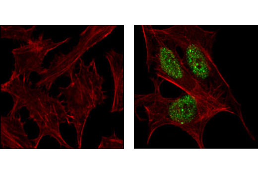 Confocal immunofluorescent analysis of HeLa cells, untreated (left) or UV-treated (right), using Phospho-53BP1 (Ser1778) Antibody (green). Actin filaments have been labeled with Alexa Fluor® 555 phalloidin (red).
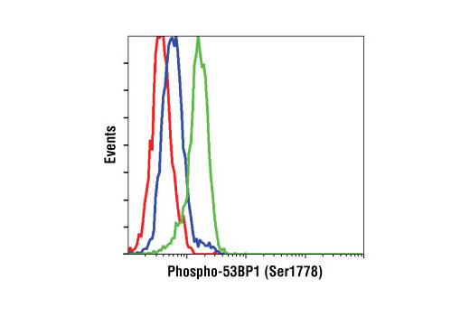 Flow cytometric analysis of HeLa cells, untreated (blue) or UV-treated (green), using Phospho-53BP1 (Ser1778) Antibody compared with a nonspecific negative control antibody (red).