53BP1 Antibody
| Name | 53BP1 Antibody |
|---|---|
| Supplier | Cell Signaling Technology |
| Catalog | 4937 |
| Prices | $246.00 |
| Sizes | 100 µl (10 western blots) |
| Host | Rabbit |
| Clonality | Polyclonal |
| Applications | WB IHC-P ICC/IF |
| Species Reactivities | Human, Monkey |
| Antigen | Polyclonal antibodies are produced by immunizing animals with a synthetic peptide corresponding to residues near the center of human 53BP1 |
| Description | Rabbit Polyclonal |
| Gene | TP53BP1 |
| Supplier Page | Shop |
Product images
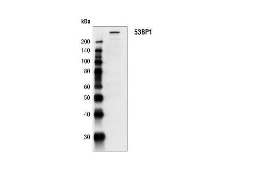 Western blot analysis of extracts from HT29 cells, using 53BP1 Antibody.
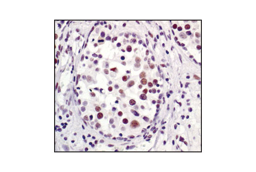 Immunohistochemical analysis of paraffin-embedded human breast carcinoma, showing nuclear localization, using 53BP1 Antibody.
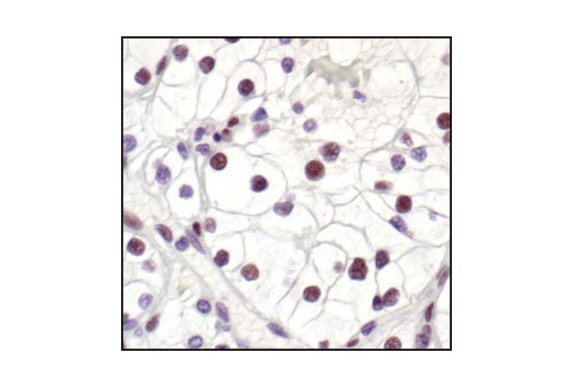 Immunohistochemical analysis of paraffin-embedded human renal cell carcinoma, using 53BP1 Antibody.
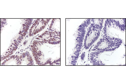 Immunohistochemical analysis of paraffin-embedded human colon carcinoma, using 53BP1 Antibody in the presence of control peptide (left) or antigen-specific peptide (right).
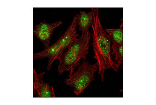 Confocal immunofluorescent analysis of HeLa cells using 53BP1 Antibody (green). Actin filaments have been labeled with Alexa Fluor® 555 phalloidin (red).