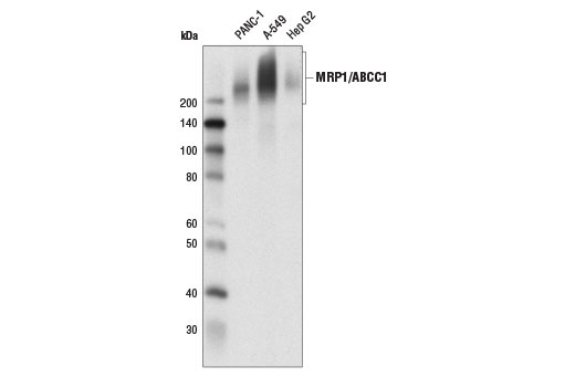
Western blot analysis of extracts from A-549, PANC-1, and Hep G2 cells using MRP1/ABCC1 (D7O8N) Rabbit mAb.
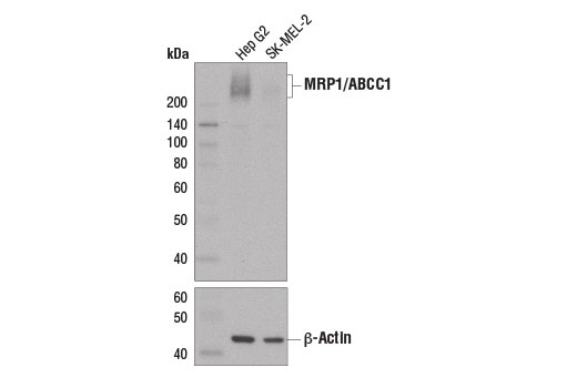
Western blot analysis of extracts from Hep G2 and SK-MEL-2 cells using MRP1/ABCC1 (D7O8N) Rabbit mAb (upper) and β-Actin (13E5) Rabbit mAb #4970 (lower).
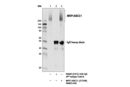
Immunoprecipitation of MRP1/ABCC1 from Hep G2 cell extracts using Rabbit (DA1E) mAb IgG XP ® Isotype Control #3900 (lane 2) or MRP1/ABCC1 (D7O8N) Rabbit mAb (lane 3). Lane 1 is 10% input. Western blot analysis was performed using MRP1/ABCC1 (D7O8N) Rabbit mAb.
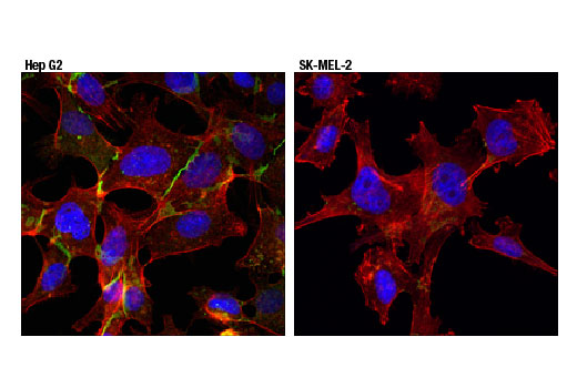
Confocal immunofluorescent analysis of Hep G2 (left) and SK-MEL-2 (right) cells using MRP1/ABCC1 (D7O8N) Rabbit mAb (green). Actin filaments were labeled with DyLight™ 554 Phalloidin #13504 (red). Blue pseudocolor = DRAQ5 ® #4084 (fluorescent DNA dye).